Anatomy of the Spinal Cord & Brain | Spinal Cord
Objective 5
Discuss and illustrate the key features of the spinal cord.
Recall that the central nervous system is comprised of the brain and spinal cord. We’ll consider the brain later, but first we will study the spinal cord.
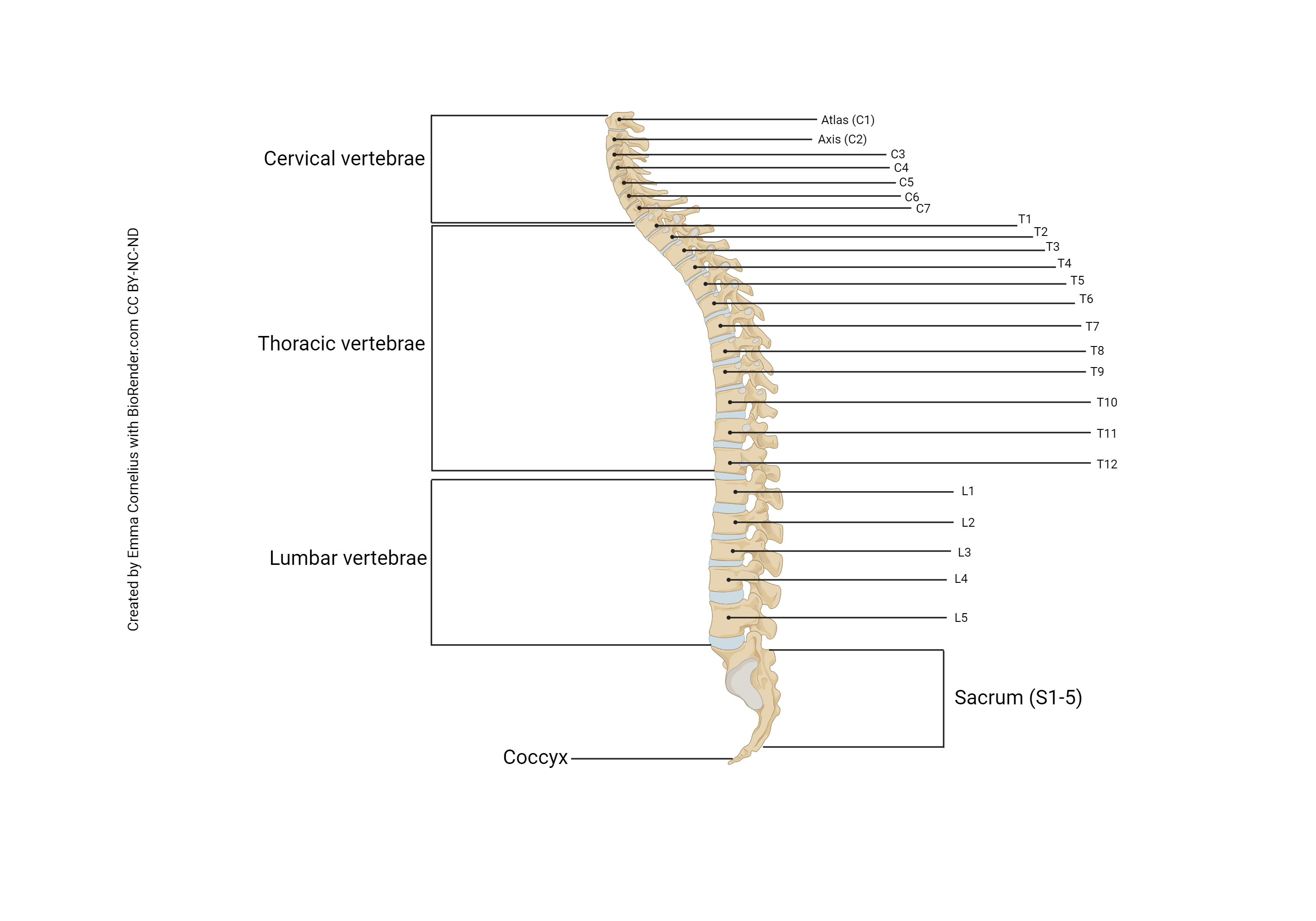
Take time now to review the anatomy of the vertebral column (Unit 9). Recall that there are 7 cervical vertebrae, 12 thoracic vertebrae, 5 lumbar vertebrae, 5 fused sacral vertebrae, and 3-4 fused coccygeal vertebrae. The vertebrae are numbered from top to bottom, with a prefix indicating cervical (C1-C7), thoracic (T1-T12), lumbar (L1-L5), or sacral (S1-S5). These will surround the spinal cord, but neurologists also define spinal cord segments which bear a relationship to the vertebrae nearby. We’ll get into the details of this relationship in Unit 12.
The spinal cord is about the diameter of your little finger at its largest (where it emerges from the brainstem at the level of the foramen magnum) and tapers to a point at its end (the filum terminale, “terminal thread”).
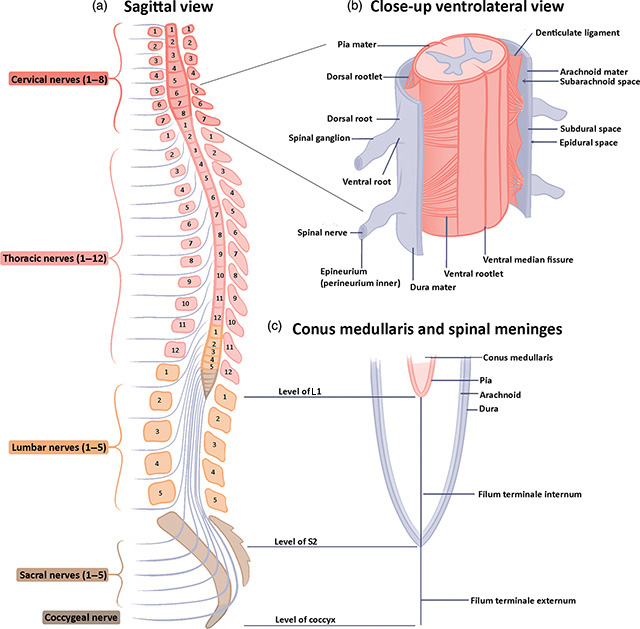
The spinal cord ends at the level of the second lumbar vertebra (L2). From there, it forms a bundle of nerves called the cauda equina (“horse’s tail”), which floats in a bag of CSF. This allows the clinician to withdraw CSF in a procedure called a lumbar puncture. Typically, lumbar punctures are done by inserting a large needle through the intervertebral disc at the L3/L4 level. In the image shown here, the spinal cord is divided into numbered segments (dark red: C1-C8; light red: T1-T12; orange: L1-L5; sacral, brown). Note the mismatch between the origin of nerve axons (in the gray matter of the spinal cord) and their emergence from the vertebral column. For example, the motor neurons which lie in the anterior horn gray matter of L5 have to travel several centimeters inferiorly to emerge below the L5 vertebra.
This also raises the issue of numbering of nerve roots. For cervical levels, the spinal cord segment and spinal nerve are named after the vertebra below the nerve. That is, the C1 spinal nerve emerges from the space between the occipital bone and the atlas (C1): remember from Unit 9 that this is called the atlanto-occipital joint. The C2 spinal nerve emerges from the joint between the atlas (C1) and the axis (C2): the atlanto-axial joint. There is an eighth cervical segment of the spinal cord (C8) but no corresponding vertebra; the C8 spinal nerve emerges from the space between the C7 and T1 vertebrae.
Then we switch to a different system. From T1 to coccygeal levels, the spinal nerve is named for the vertebra above where the nerve emerges. Thus the posterior (dorsal) horn of the T1 segment of spinal cord receives sensory information carried on part of the T1 spinal nerve. The T1 segment of the anterior (ventral) horn has α motor neurons that send information on axons that make up part of the T1 spinal nerve. The T1 spinal nerve emerges from below the T1 vertebra. The T2 spinal nerve emerges from below the T2 vertebra, and so forth.
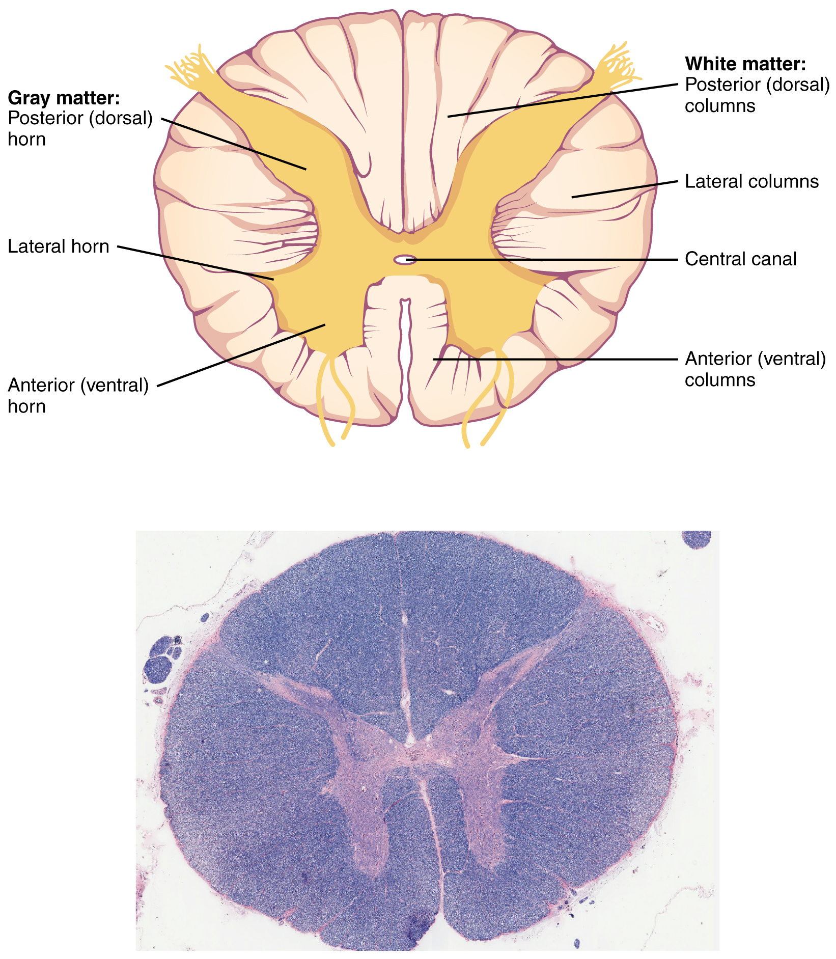
In cross-section, the spinal cord is oval or round and consists of an H– or butterfly-shaped central core of gray matter surrounded by white matter (i.e., myelinated axons traveling together over a great distance). Posterior (dorsal) roots of spinal nerves bring sensory information into the spinal cord. Anterior (ventral) roots of spinal nerves carry motor information away from the spinal cord.
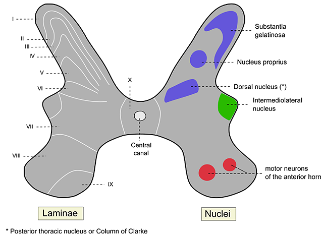
The H-shaped gray matter is further subdivided into a posterior (dorsal) horn, which processes the sensory information brought in by the posterior root; and an anterior (ventral) horn, which contains cell bodies of α motor neurons (alpha motor neurons) controlling muscles. The axons of the α motor neurons form the anterior root of the spinal nerve, which carries motor commands going out to muscles.
At thoracic and upper lumbar levels, there is also a lateral horn (also called the intermediolateral nucleus or column) which contains the cell bodies of the autonomic neurons we saw in Objective 2. These will be the focus of Unit 12 Objective 2. At thoracic levels, these are the cell bodies of the sympathetic neurons. These are called preganglionic neurons and they send a short axon to the nearby sympathetic chain ganglia where they synapse onto postganglionic neurons which send a long axon to reach the actual organ being innervated. For example, this is how the heart speeds up when you activate the sympathetic nervous system. (There is a diagram of this in Unit 12 and in Unit 16.)
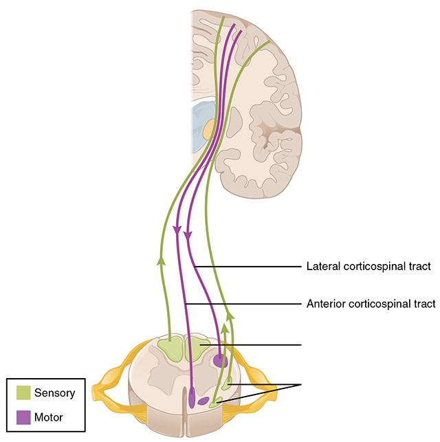 The white matter of the spinal cord is on the outside (superficial). It contains the axons of neurons that are either carrying information from the cerebral cortex of the brain to the spinal cord (called corticospinals) and from the brainstem to the spinal cord. Collectively, these are called descending tracts. There are also axons of nerve cells carrying information from the body, or from the spinal cord, to the brain. These are collectively called ascending tracts.
The white matter is further subdivided into three zones that are named pretty logically. The posterior (dorsal) horn gray matter extends to the surface and forms the “walls” of a posterior column. In the lateral white matter is the lateral column. The anterior (ventral) white matter is the anterior column.
Media Attributions
- U11-045 Spinal Cord Cross Section Diagram © Betts, J. Gordon; Young, Kelly A.; Wise, James A.; Johnson, Eddie; Poe, Brandon; Kruse, Dean H. Korol, Oksana; Johnson, Jody E.; Womble, Mark & DeSaix, Peter is licensed under a CC BY (Attribution) license
- U11-047 Vertebral Column © Cornelius, Emma is licensed under a CC BY-NC-ND (Attribution NonCommercial NoDerivatives) license
- U11-047 Spinal_cord_details corrected © Sheryl Tan, Richard LM Faull, Maurice A Curtis is licensed under a CC BY (Attribution) license
- U11-048 rexed laminae and functional regions of gray matter © German version by Polarlys, translated by Interiot is licensed under a CC BY (Attribution) license
- U12-010 Spinal Cord Pathways Motor Focus v2 © Betts, J. Gordon; Young, Kelly A.; Wise, James A.; Johnson, Eddie; Poe, Brandon; Kruse, Dean H. Korol, Oksana; Johnson, Jody E.; Womble, Mark & DeSaix, Peter adapted by Jim Hutchins is licensed under a CC BY (Attribution) license
