Anatomy of the Spinal Cord & Brain | Posterior Root Ganglion & Spinal Nerves
Objective 6
Indicate the position of the posterior root ganglion and spinal nerves.
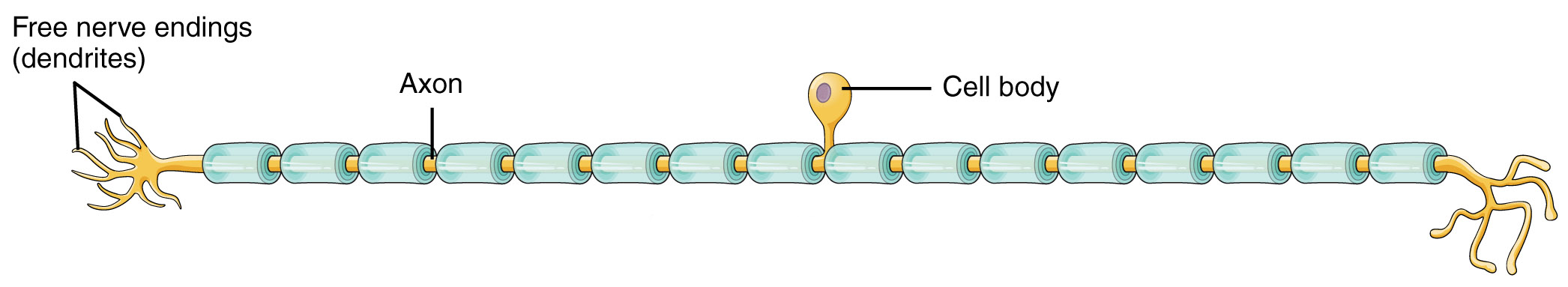
Remember that in Objective 3 we studied three basic shapes of neurons. One of these, the unipolar neuron, is found in skin sensory pathways. Modified dendrites are found in the dermis of the skin. They decide whether to create an electrical signal (action potential) within the dermis; if an action potential is created, it travels without modification along a long axon within a spinal nerve and into the posterior horn of the spinal cord directly. These axons also form the posterior roots of the spinal nerve.
Note that the previous paragraph described modified dendrites and an axon, but did not mention the cell body. Where is the cell body? It is located in a posterior root ganglion, a collection of nerve cell bodies found in the PNS. The posterior root ganglion (also called a dorsal root ganglion) is found just outside of the vertebral column and is therefore not covered by bone like the CNS structures are.
Motor information is processed and sent by α motor neurons whose axons travel out the anterior root.
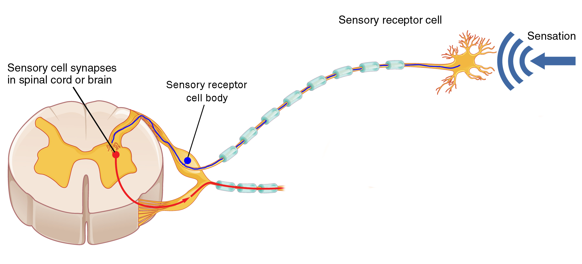
Just outside the enclosure of the vertebrae, the posterior root and anterior root join anatomically to form a spinal nerve. (Notice that the information in the spinal nerve is traveling in both directions: sensory information entering the CNS via the posterior root, and motor information leaving the CNS via the anterior root.)
As we saw in Objective 2, there is a different anatomical setup for the sympathetic nervous system. Autonomic motor neurons are found in the lateral horn from thoracic level 1 (T1) to lumbar level 2 (L2).
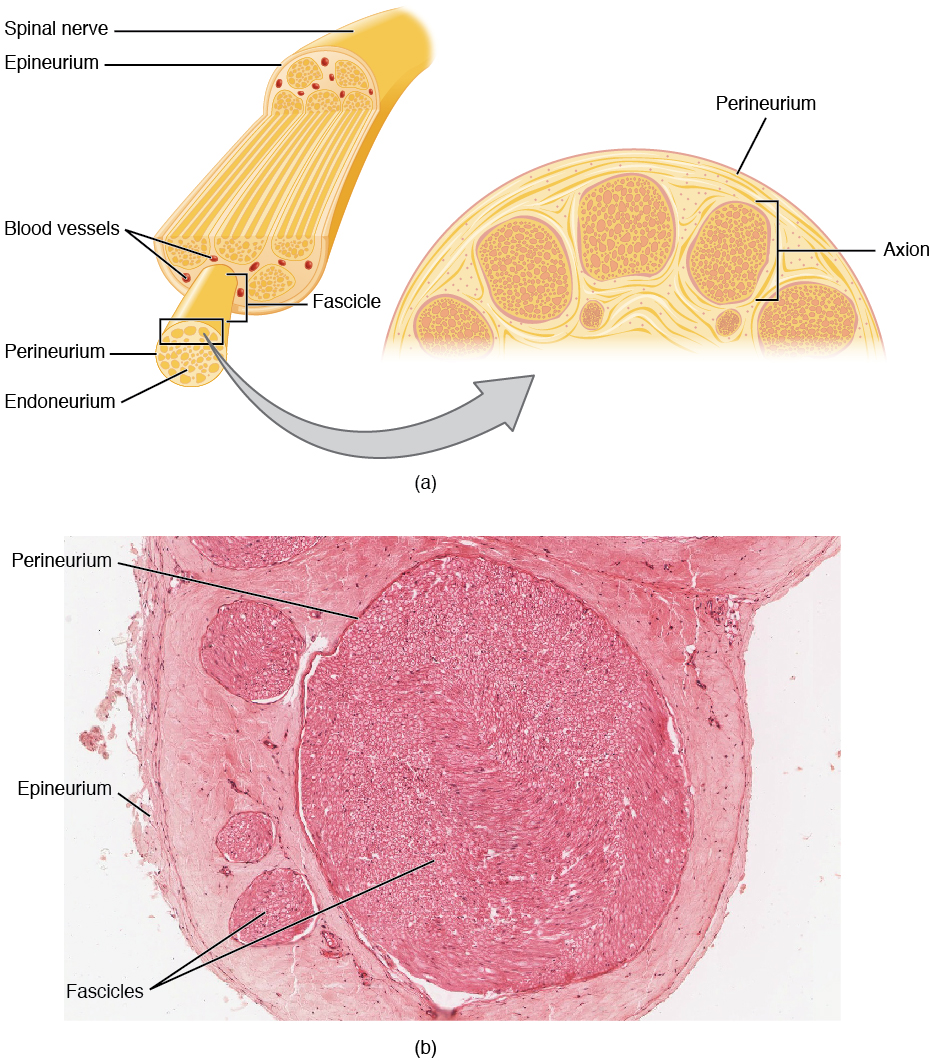
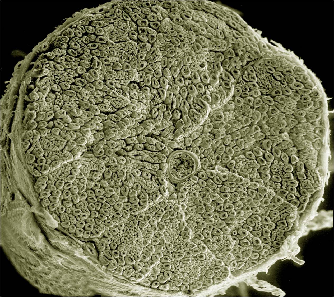
The microscopic structure of spinal nerves is shown in this diagram and scanning electron micrograph. As we’ve seen previously, spinal nerves are mixed: they contain motor neuron axons (both somatic and autonomic) as well as sensory neuron axons. Individual axons may be wrapped by Schwann cells with a myelin sheath (myelinated). Alternatively, they might be covered by a Schwann cell without being wrapped in myelin (unmyelinated).
As with muscle, there are three types of connective tissue sheath around nerve axons.
- endoneurium surrounds individual myelinated axons;
- perineurium surrounds bundles of axons, called fascicles;
- epineurium surrounds the entire nerve.
At several places in the body, spinal nerve roots and spinal nerves swap axons around so they don’t always take straight line paths. Because these structures resemble a braid, they have the Latin name for braid: plexus (plural plexi).
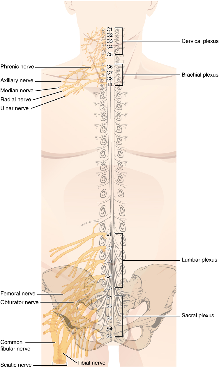 There are, in all, four of these braided nerve structures associated with the spinal cord.
- The cervical plexus is in the neck. It receives nerves from the C1 through C5 spinal cord segments, and gives rise to several important nerves. The most important of these is the phrenic nerve, which innervates the diaphragm and makes breathing possible. Its contributory nerves are C3, C4 and C5, thus the mnemonic “C3, 4, 5, keep the diaphragm alive.”
- The brachial plexus receives contributions from C5-C8 and T1. Its major exiting nerves are the radial nerve (innervating the thumb and nearby structures), the median nerve (middle finger) and ulnar nerve (little finger). The nerves fight over the index and ring fingers.
- The lumbar plexus is associated with the lumbar spinal cord (roots L1–L5) and innervates the upper thigh.
- The sacral plexus receives contributions from L4–L5 and S1–S5. One huge nerve, the largest in the body, emerges from the sacral plexus: the sciatic nerve. Almost every axon innervating muscles in the leg, and almost all sensory information from the leg, is carried by the sciatic nerve. Imagine how painful inflammation or compression of this nerve would be, a condition called sciatica.
1 There are seven cervical vertebrae and eight cervical nerves. C1 nerve comes out above C1 vertebra, between the occiput of the skull and the atlas (C1 vertebra). C2 nerve comes out above C2 vertebra (axis), and so forth, until C7 nerve, which emerges from above C7 vertebra. Then we have C8 nerve, which emerges from below C7 vertebra. T1 nerve comes out below T1 vertebra, and from there down, that’s how the nerves are named: by the vertebra above.
Media Attributions
- U11-049 Sensory Receptors © Betts, J. Gordon; Young, Kelly A.; Wise, James A.; Johnson, Eddie; Poe, Brandon; Kruse, Dean H. Korol, Oksana; Johnson, Jody E.; Womble, Mark & DeSaix, Peter adapted by Jim Hutchins is licensed under a CC BY (Attribution) license
- U11-050 Posterior Root Ganglion © Betts, J. Gordon; Young, Kelly A.; Wise, James A.; Johnson, Eddie; Poe, Brandon; Kruse, Dean H. Korol, Oksana; Johnson, Jody E.; Womble, Mark & DeSaix, Peter adapted by Jim Hutchins is licensed under a CC BY (Attribution) license
- U11-051 Spinal Nerve © Betts, J. Gordon; Young, Kelly A.; Wise, James A.; Johnson, Eddie; Poe, Brandon; Kruse, Dean H. Korol, Oksana; Johnson, Jody E.; Womble, Mark & DeSaix, Peter is licensed under a CC BY (Attribution) license
- U11-052 Spinal Nerve Human © Ellisman, Mark and Deerinck, Tom, National Center for Microscopy and Imaging Research is licensed under a Public Domain license
- U11-054 Nerve Plexuses © Betts, J. Gordon; Young, Kelly A.; Wise, James A.; Johnson, Eddie; Poe, Brandon; Kruse, Dean H. Korol, Oksana; Johnson, Jody E.; Womble, Mark & DeSaix, Peter is licensed under a CC BY (Attribution) license
