Anatomy of the Spinal Cord and Brain | Cerebrum, Cerebellum, and Brainstem
Objective 7
List and label the most important structures of the brain.
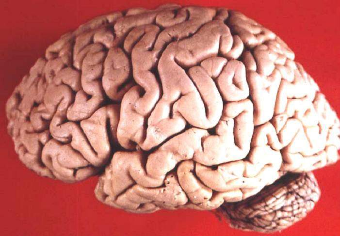
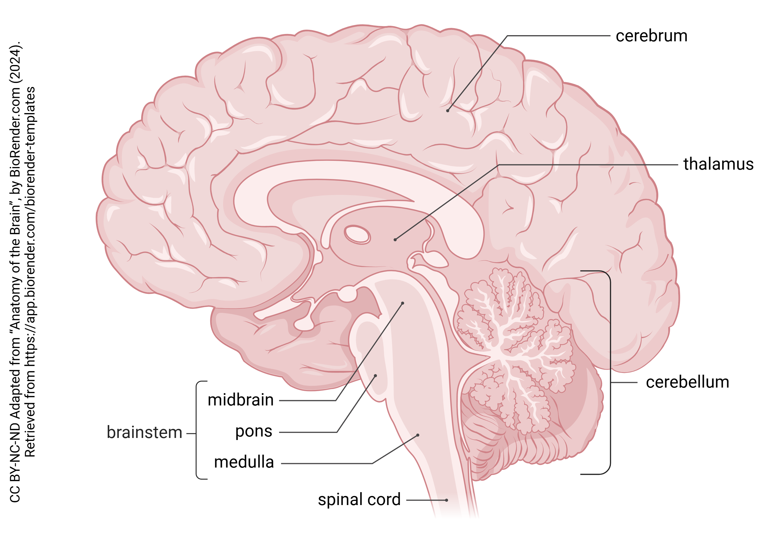
This is the human brain in lateral view. In order to understand its anatomy, we have to understand how such a complex and elaborate structure develops.
The central nervous system of the human begins to develop shortly after gastrulation, the formation of three primordial layers that occurs 16 days post-conception (30 days since the last period). A part of the future skin (ectoderm) begins to thicken, forming a neural plate. The plate then develops a shallow groove that deepens into a neural fold over the next few days.
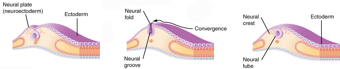 The edges of this groove fold up and seal together forming a neural tube. This tube then begins to change shape and develop empty rooms called vesicles. The first set of three vesicles are called primary brain vesicles. The second set of vesicles are called secondary brain vesicles and we need to learn the names of these because those will help us understand the anatomy of the adult nervous system.
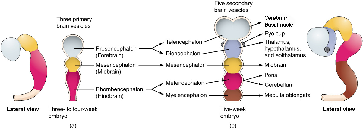
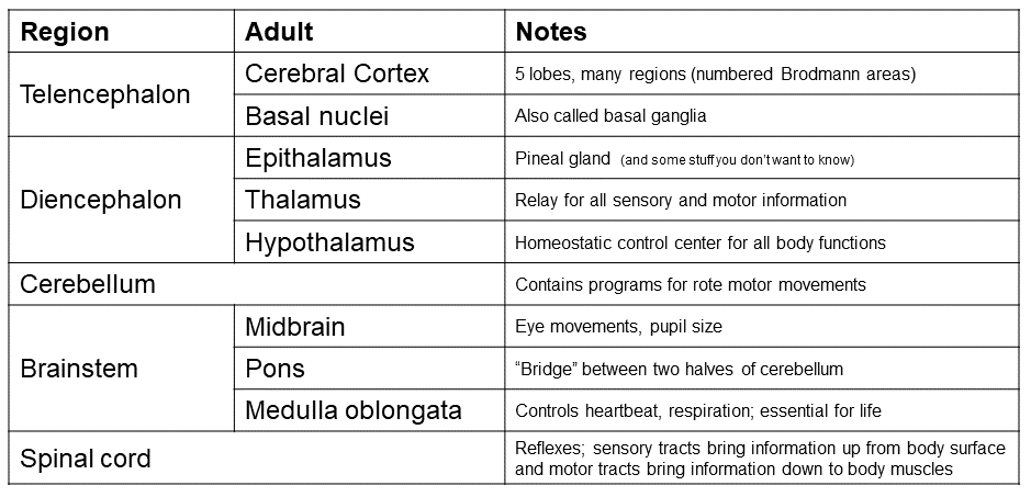
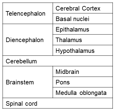 These subdivisions of the developing brain become adult structures, as shown in the table. Because each of these areas has a different anatomy and different strategy of “brain wiring”, it’s important to understand just a little bit about the development of the brain to understand the anatomy and function of the brain.
For example, the cerebral cortex and basal nuclei, which represent the vast majority of the weight of the human brain, are extensively interconnected and control much of human behavior. The basal nuclei smooth out the activity of the part of cerebral cortex that controls muscles; this smoothing goes wrong in Parkinson disease. Changes in the activity of another part of the basal nuclei is implicated in obsessive-compulsive disorder. We will examine some of the signaling molecules and wiring of this system in Unit 12, and we will describe what goes wrong with signaling in the basal nuclei in the Pathophysiology course.
(Many authorities call the basal nuclei the basal ganglia. I believe this is confusing; a ganglion, as we’ve just discussed, is a collection of nerve cell bodies in the PNS. The basal nuclei fit squarely within the CNS. That’s a good reason to go through the hassle of changing the name for our sons and daughters who might have to learn neuroanatomy later.)
This table contains the names of all the brain structures we need to learn at this point in the course.
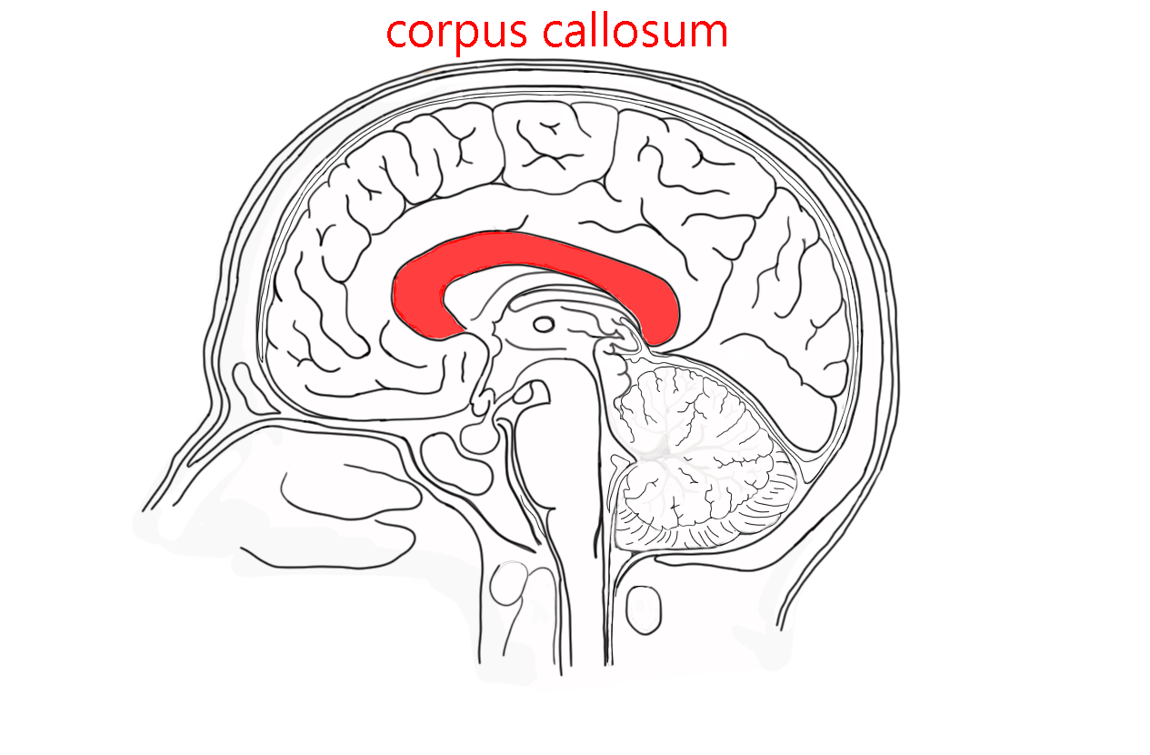
A deep groove, the medial longitudinal fissure, divides the brain into a left hemisphere and right hemisphere. These are connected by the corpus callosum, a band of axons running between mirror-image areas of the brain, right and left.

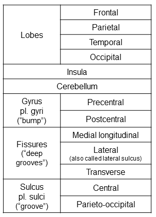
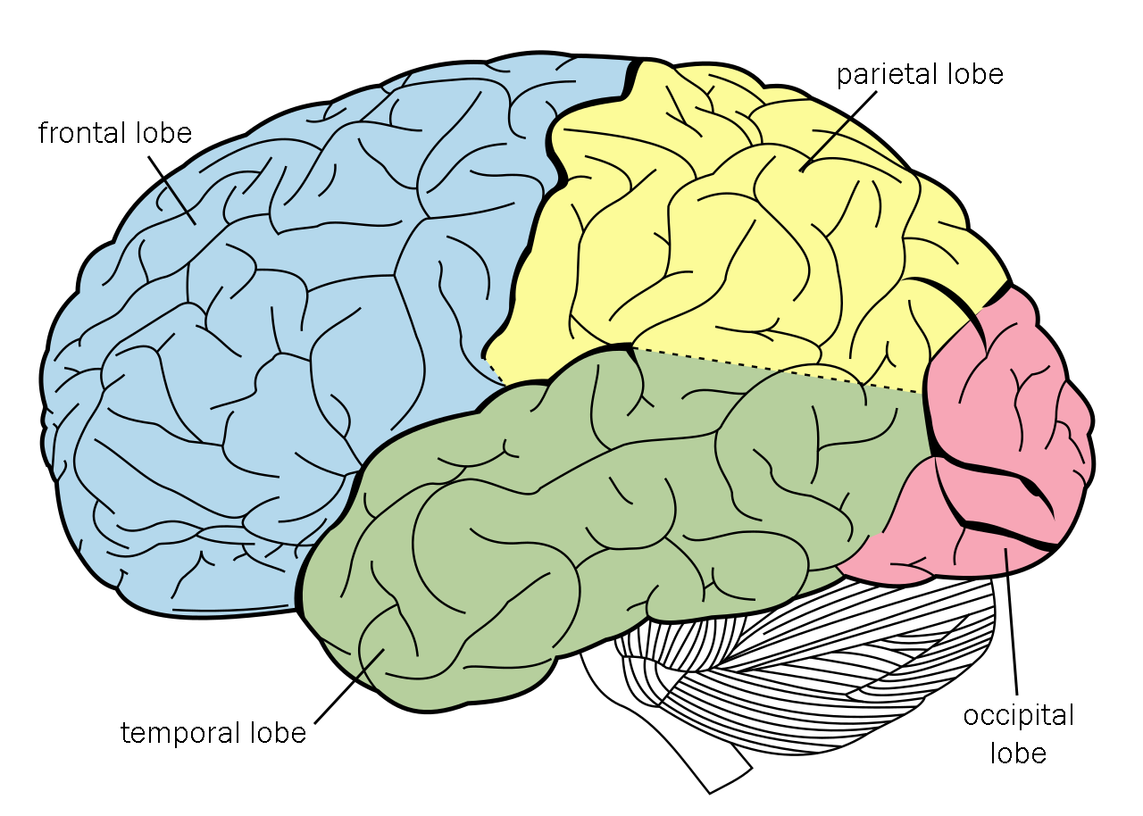
First, we’ll divide the external surface of the cerebral cortex into four lobes.
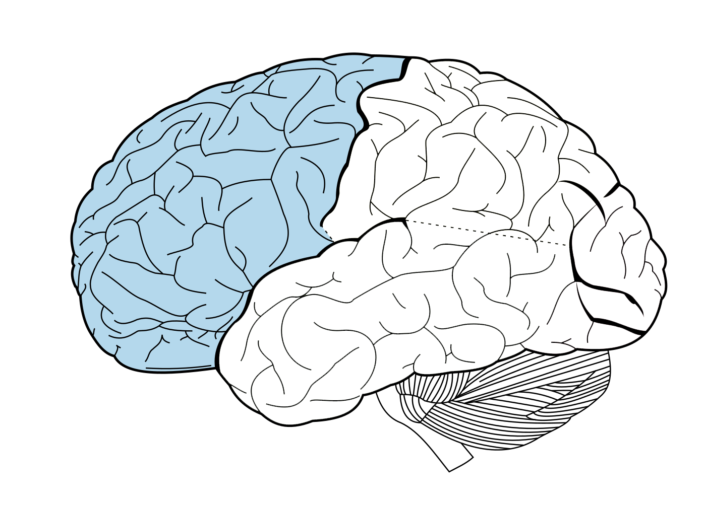
The frontal lobe (wait for it…) is in front. (left)
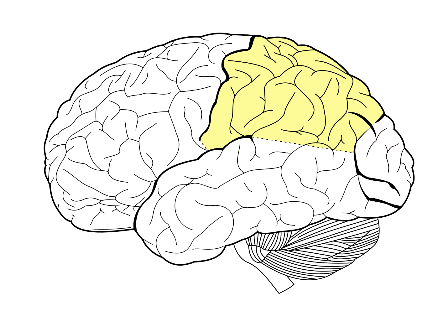
The parietal lobe shares its borders with the other three external lobes. It is just posterior to the frontal lobe. (right)
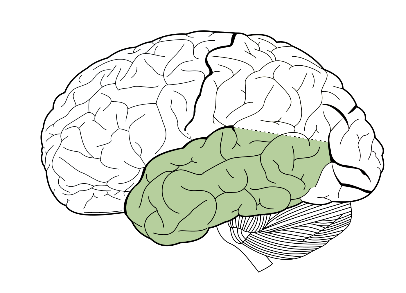
The temporal lobe (wait for it…) is under the temple. (left)
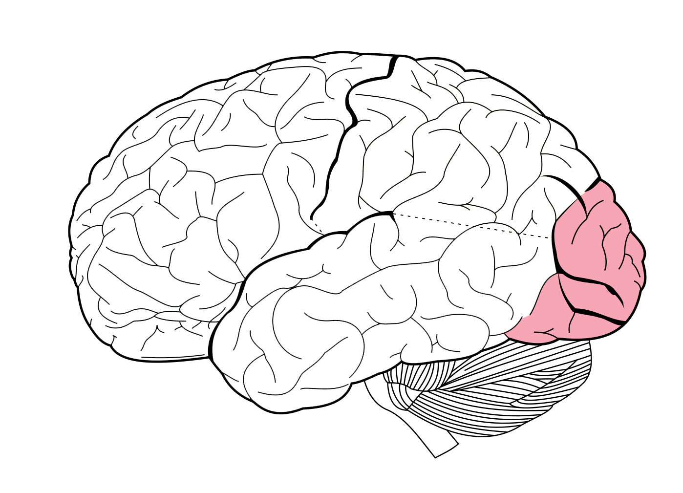
The occipital lobe is the most posterior lobe of the brain. (right)
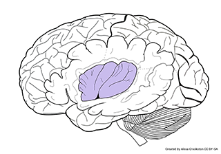 The insula is a fifth lobe (insular lobe). This is the part of the brain that is folded inside, and only visible when we cut the lateral surface of the brain away. Scientists believe the insula is the source of social emotions like guilt or pride. It may also be the area of the brain that interprets or drives cravings.
The cerebellum, attached to the brainstem, is a separate cortex (cerebellar cortex) with a separate structure.
Its job is to compare the motor programs of the brain to what the body is actually doing in real time and correct any mistakes that are being made. It is connected to the brainstem by three cerebellar peduncles (“little feet”).
 Fissures or grooves in the surface of the cortex are called sulci (singular, sulcus; Latin, “furrow, groove, ditch, trench”). Deep grooves are called fissures. Bumps on the surface are called gyri (singular, gyrus; Latin: “ring, circle”).
We have already encountered the medial longitudinal fissure (sometimes called the longitudinal fissure), a deep groove which divides the brain into right and left halves.
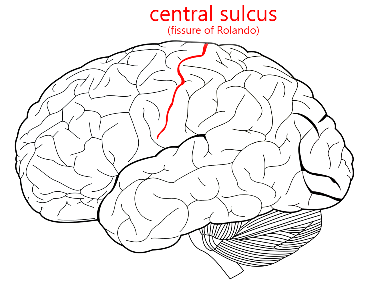
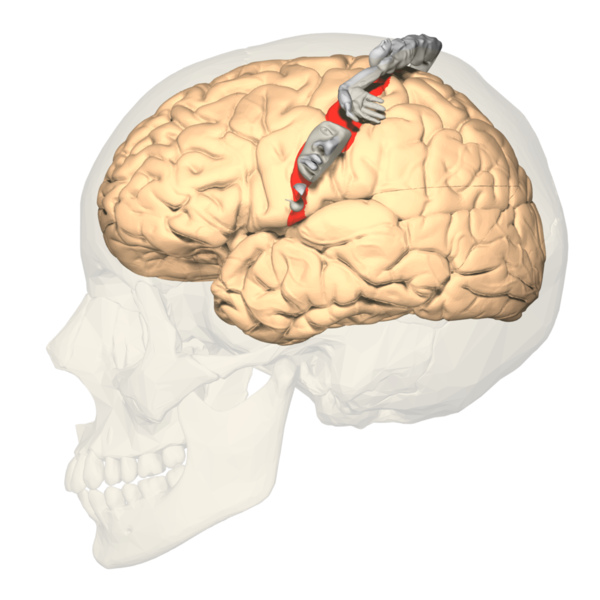
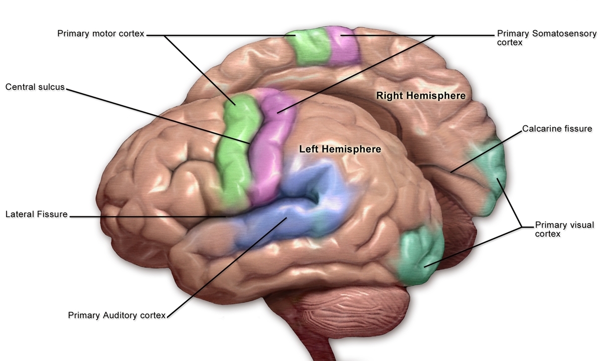
The frontal lobe and parietal lobe are separated by the central sulcus. The name seems to say that it’s easy to locate; in practice, it’s harder to find than you might think. The central sulcus forms the border between the frontal lobe and parietal lobe.
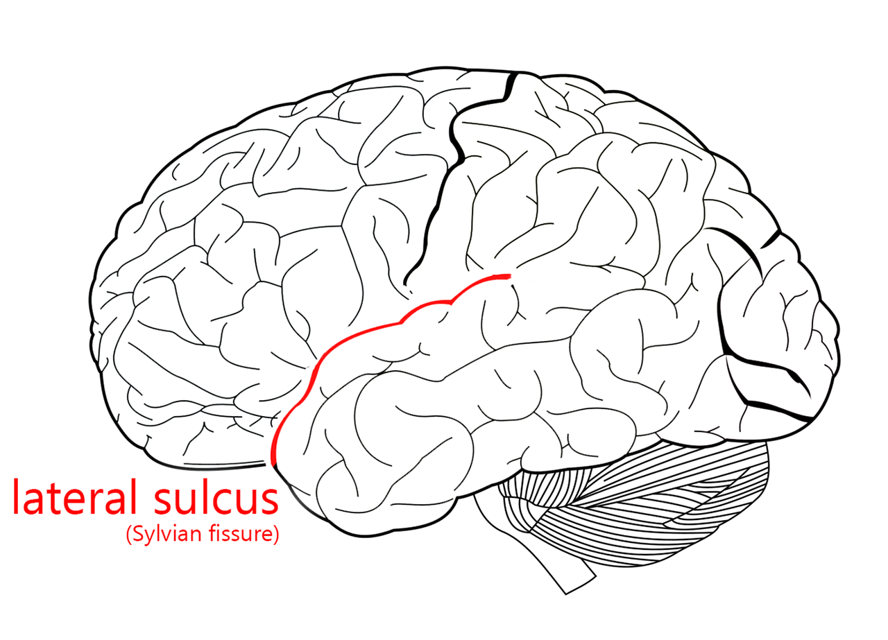
The lateral sulcus is often called the lateral fissure. As the name implies, it’s probably the most prominent feature of the lateral surface of the brain. Whenever I draw a stylized lateral view of the brain, I include the lateral sulcus and the outline of the brain and I’m done.
The lateral sulcus divides the frontal and parietal lobes superiorly from the temporal lobe inferiorly. It becomes shallower as it passes posteriorly and is surrounded by the angular gyrus, an area important in human language.
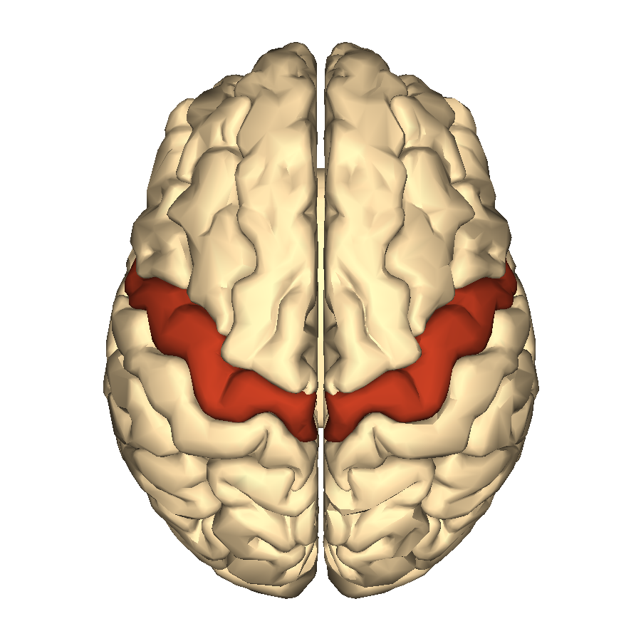
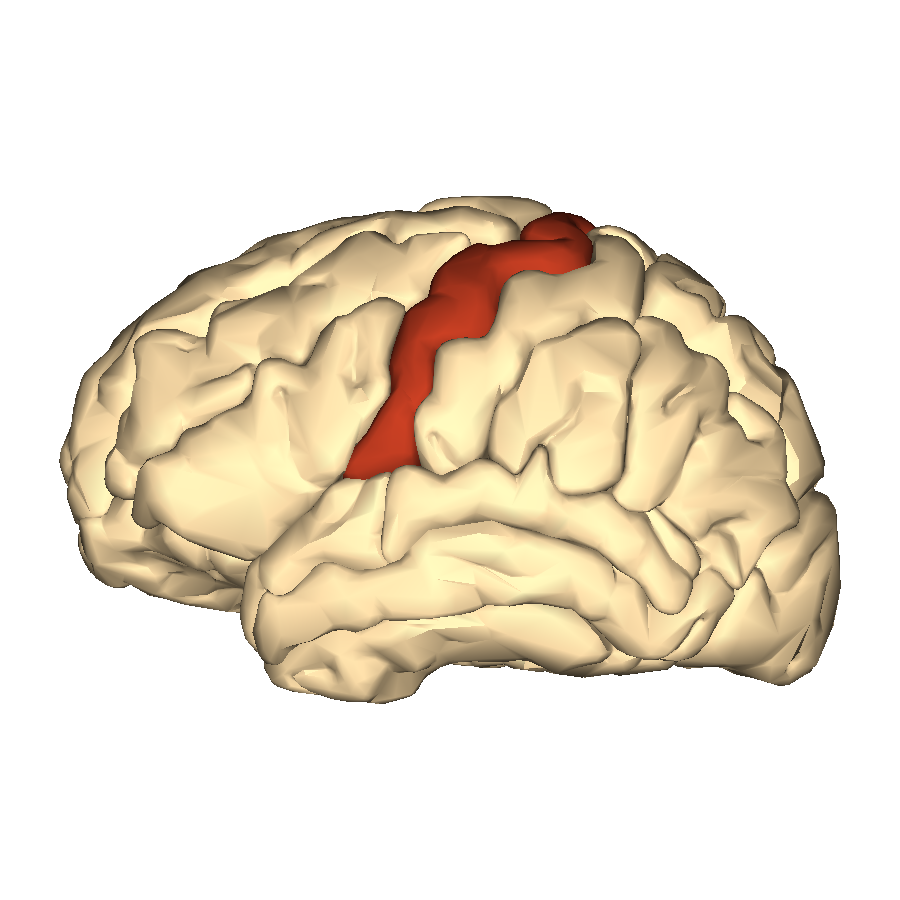
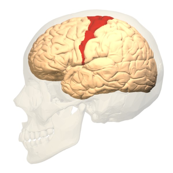
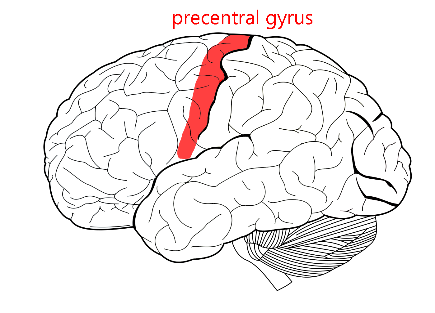 On either side of the central sulcus are two parallel, almost identical-looking gyri. The first we’ll look at is the precentral gyrus (part of the frontal lobe). As we’ll see in Unit 12, the precentral gyrus is involved in the motor system, which has an orderly map of the body distributed across the surface of the gyrus. If you make a conscious movement, the signal needed for each muscle is calculated in this area and then sent as a neural signal down to the α motor neuron in the anterior horn of the spinal cord. Because this pathway goes from cortex to spinal cord, it’s called the corticospinal tract.
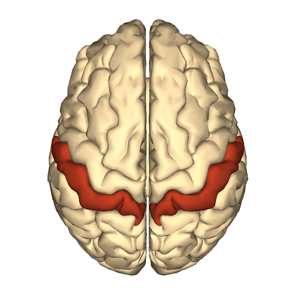
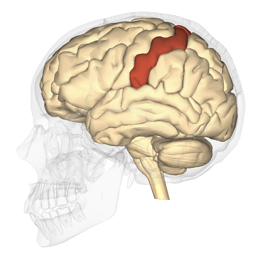
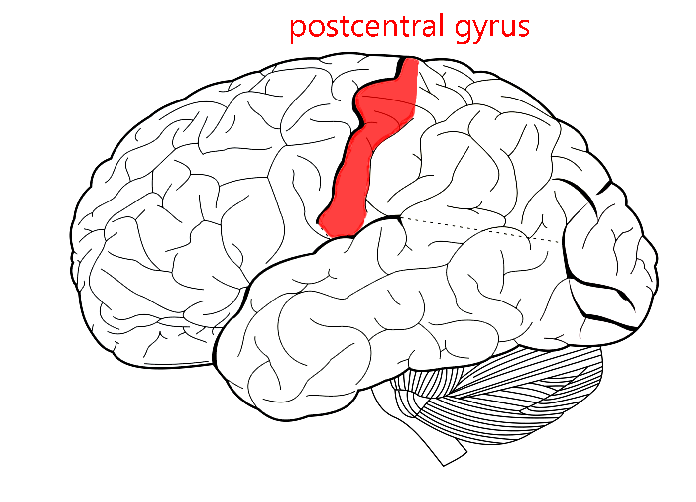 The postcentral gyrus processes sensations from the face and body surface (the somatosensory system). Like the motor cortex, it has an orderly map (homunculus) which is shown here.
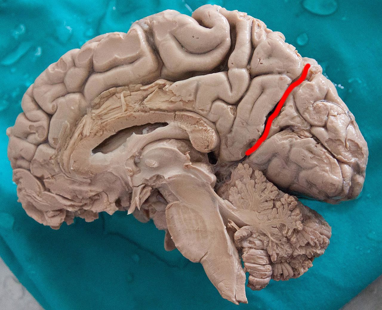
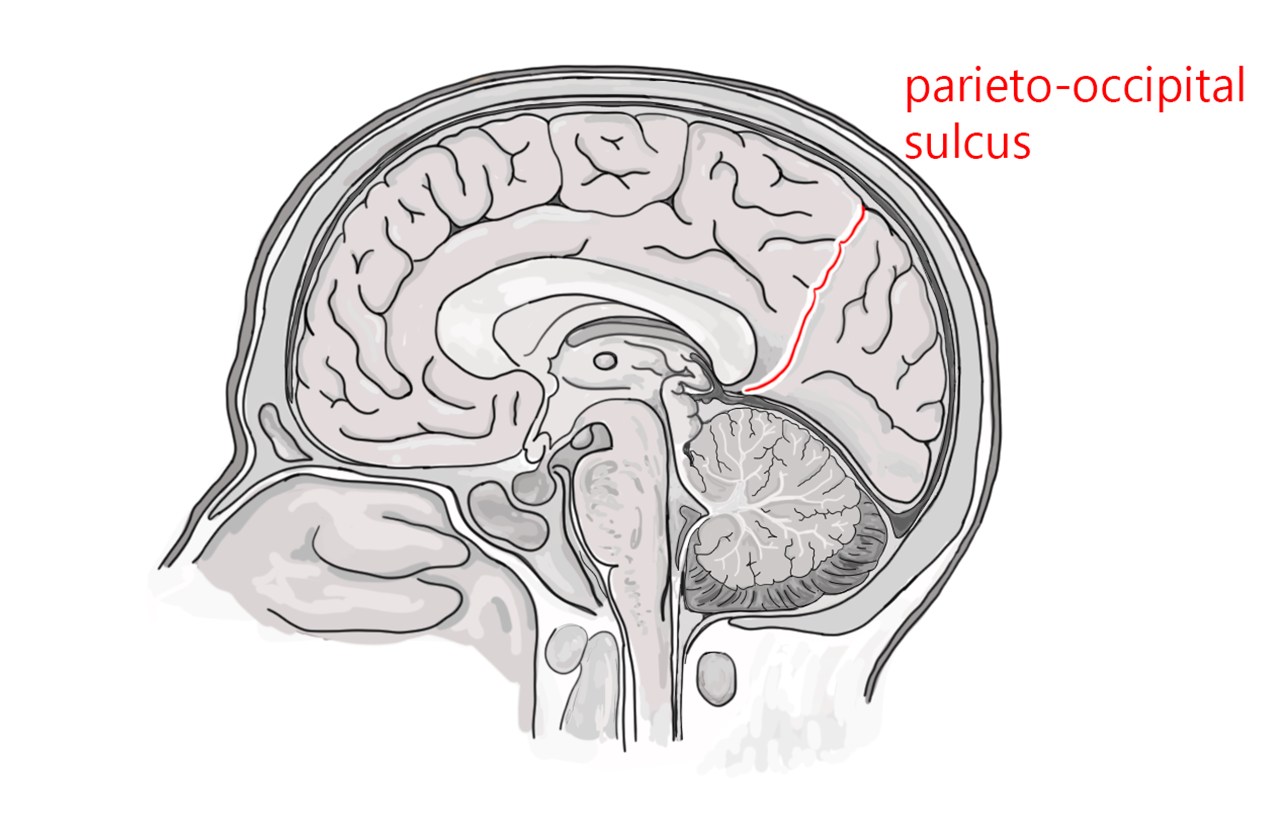
The parietal lobe and occipital lobe are separated by the parieto-occipital sulcus. The good news is, it’s a reasonable name for a sulcus separating the parietal and occipital lobes.
The bad news is, it’s only visible on the midsagittal surface.
Some authorities include the thalamus as part of the brainstem; others consider the thalamus to sit atop the brainstem. Either way, the thalamus is an area that relays information going up to cortex, and relays information leaving cortex on its way to the brainstem or spinal cord.
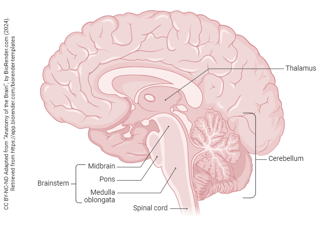
The brainstem is divided into three regions and a sort-of annex. From rostral (top) to caudal (bottom), they are the:
- midbrain
- pons
- medulla oblongata
The midbrain is the smallest major division of the brain. The most important function of the midbrain is controlling visual reflexes, like pupil size. When we look at a patient’s pupils with a flashlight, we are assessing the function of the midbrain.
The pons (Latin: “bridge”) consists of a massive band of fibers sweeping from one side of the brainstem to the other. This means it forms a characteristic bulge on the ventral (anterior) side of the brainstem and it’s easy to spot. What are all those fibers doing? They are carrying information from one side of the cerebellum to the other. You can think of the cerebellum as a sort-of annex of the brainstem.
The medulla oblongata (usually called by her friends simply “medulla”) is where the core functions of the nervous system reside. Both heartbeat and respiration are controlled from this area, and destruction of the medulla results in death.
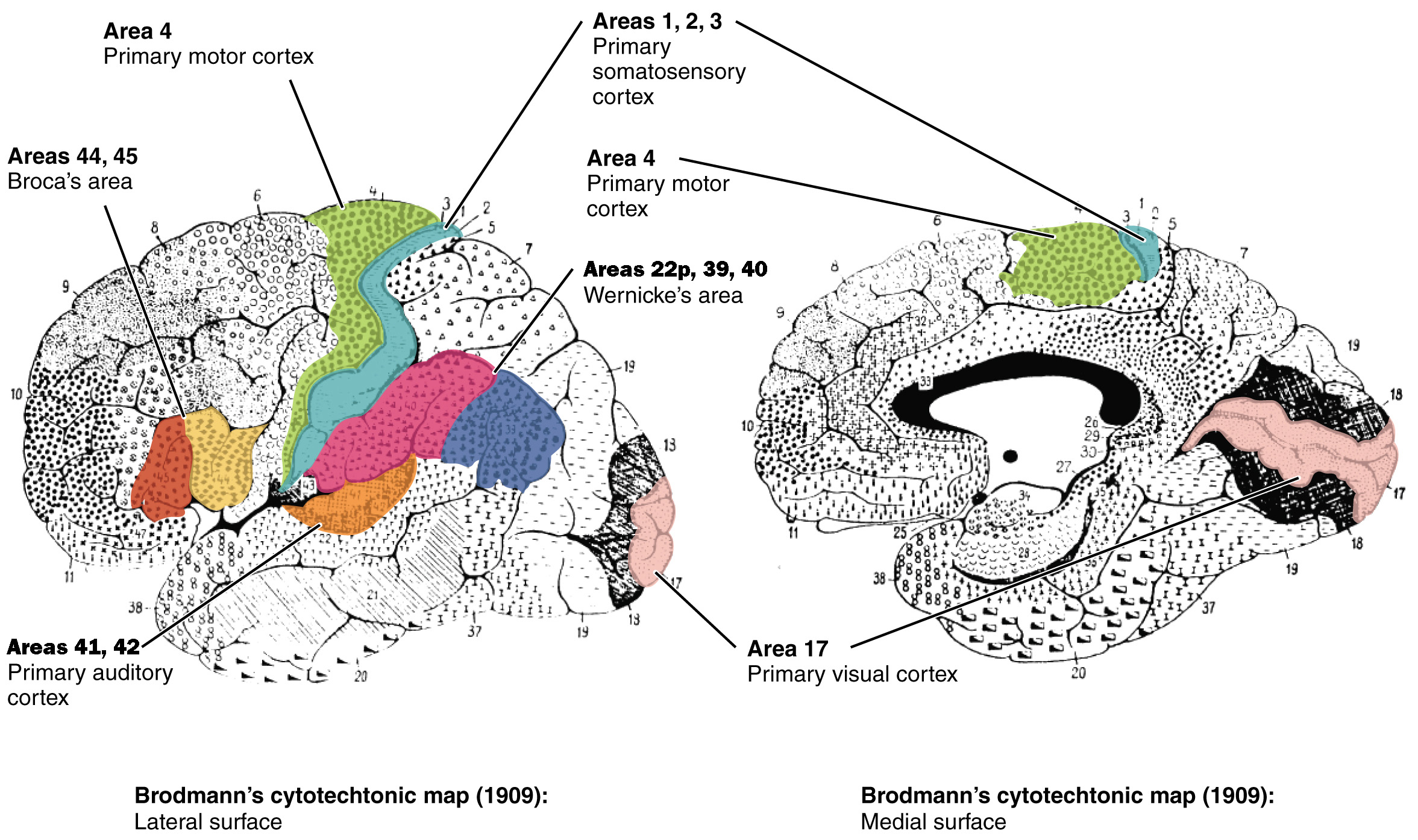
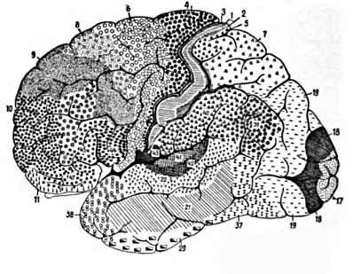
Returning to the cerebral cortex, now that we have some landmarks and lobes in mind, we can begin to further subdivide the cortex. The most common mapping system for the cortex was developed by Korbinian Brodmann in the late 1800s and early 1900s.
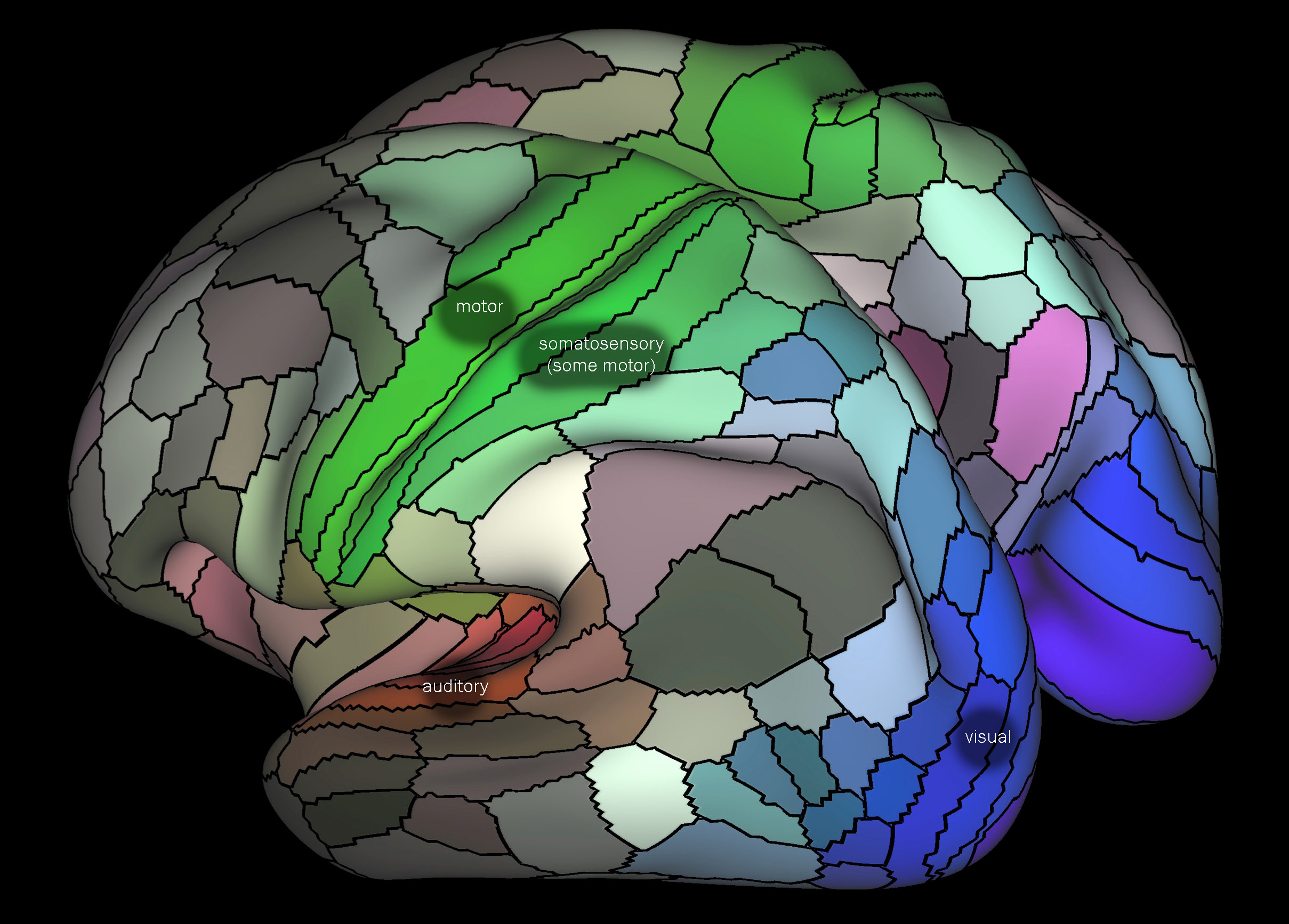
Brodmann was a German scientist who started slicing a brain and looking at the sections through a microscope. It’s surprising, as shown in the multicolored map developed in 2016 by Glasser and van Essen, how much modern maps resemble the classic map of Brodmann.
For no reason that we can fathom, Brodmann started at the top and cut horizontal sections. Most of the cerebral cortex has six layers of cells, but the way the cells are packed and the shape of the cells are different in different places. The first pattern of cell shapes and packing he named Area 1; the second, Area 2; and so forth. The last area he numbered was Area 52.2
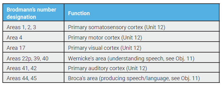
Some of the areas are synonymous with functional areas of the cortex and are still used today; others are obscure. Some of the area numbers you will frequently encounter are listed.
 Parietal Lobe
Areas 3, 1, and 2 (postcentral gyrus): primary somatosensory cortex, receiving information about the face and body surface. No one knows why we list them in this order. Taste is also here, and nearby in area 43.
2 Sadly, while in some animals there is an area 51, he skipped area 51 in humans. Maybe it’s a secret government coverup (http://skepdic.com/area51.html).

Occipital Lobe
Areas 17, 18 and 19: Visual cortex (sensory, sight). Area 17 is also called primary visual cortex (V1) or striate cortex.
Temporal Lobe
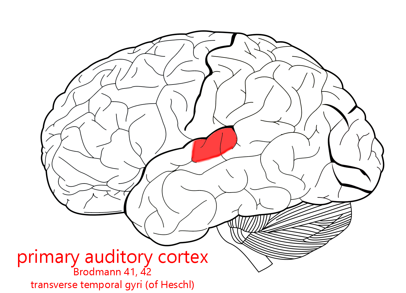 Areas 41 and 42: Primary auditory cortex (sensory, hearing).
Area 22p (posterior part of 22): Wernicke’s area. Responsible for the understanding of speech sounds.
Frontal Lobe
Area 4: (precentral gyrus): primary motor cortex, sending axons to the α motor neurons of spinal cord (executing movement).
Area 6: supplementary motor area (planning or imagining movement)
Area 8: frontal eye fields (eye movements).
Areas 44 and 45: Broca’s area. In most people, Broca’s area on the left side is responsible for the production of speech (movements of the throat and tongue).
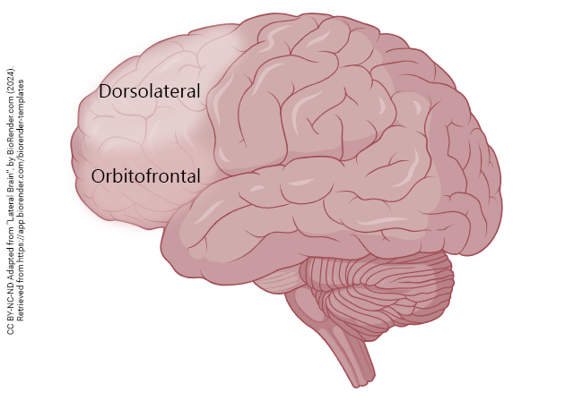 The non-motor areas of frontal cortex, in the rostralmost part of this large cortical lobe, are called prefrontal cortex. Prefrontal cortex is important for holding on to our personality and individuality, the things that make you “you”.
Among the important programs stored here are toilet training and social graces.
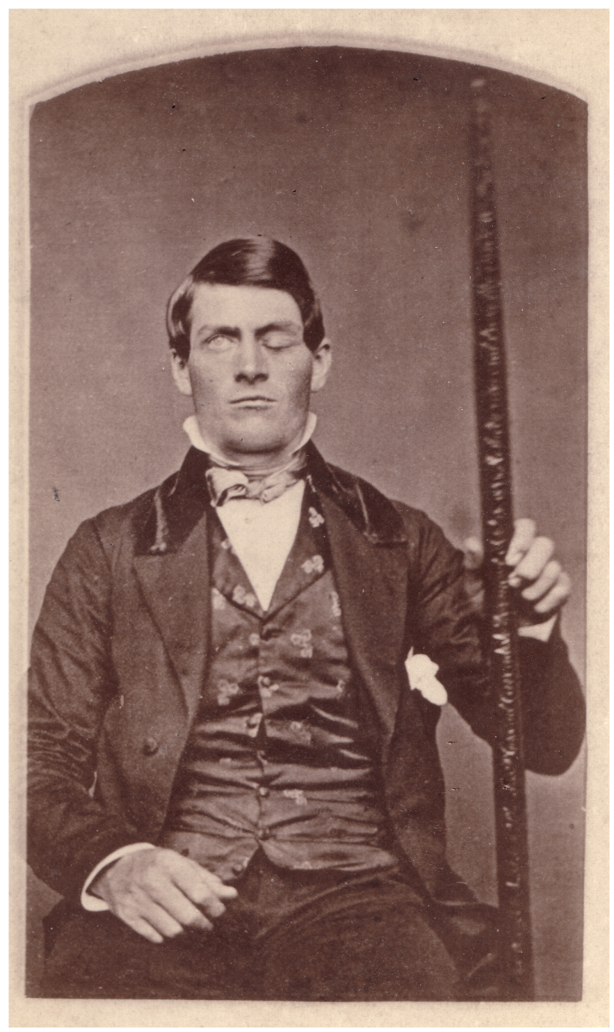
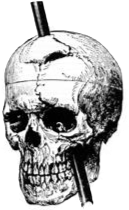
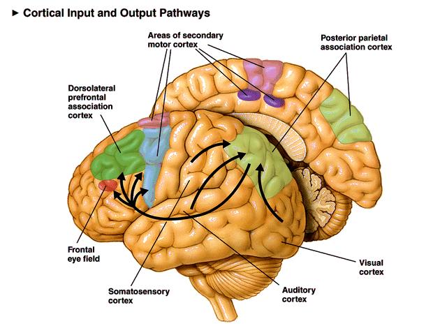
The story of how we discovered the importance of prefrontal cortex is both fascinating and horrifying. In 1848, a railroad construction foreman named Phineas Gage suffered a terrible accident: a pointed steel rod, about 4 feet long and 1 ¼ inch diameter, was blown through his left cheek and out the top of his head, obliterating his left eye and a good chunk of prefrontal cortex in the process. Miraculously, Gage survived. However, his personality changed and he was no longer much fun to be around. In essence, he lost his kindergarten training. This was the first indication that specific functions, in this case personality, could reside in specific areas of the brain. When we talk about motor cortex, or visual cortex, or auditory cortex, as we have above, it’s because Gage’s accident pointed the way to our conception of the brain as made up of a cluster of semi-independent modules each performing a specific function. This view has been validated by neurological studies (for example in stroke) that have been carried out in the almost 200 years since his accident. Some medical pioneers had no choice in the matter.
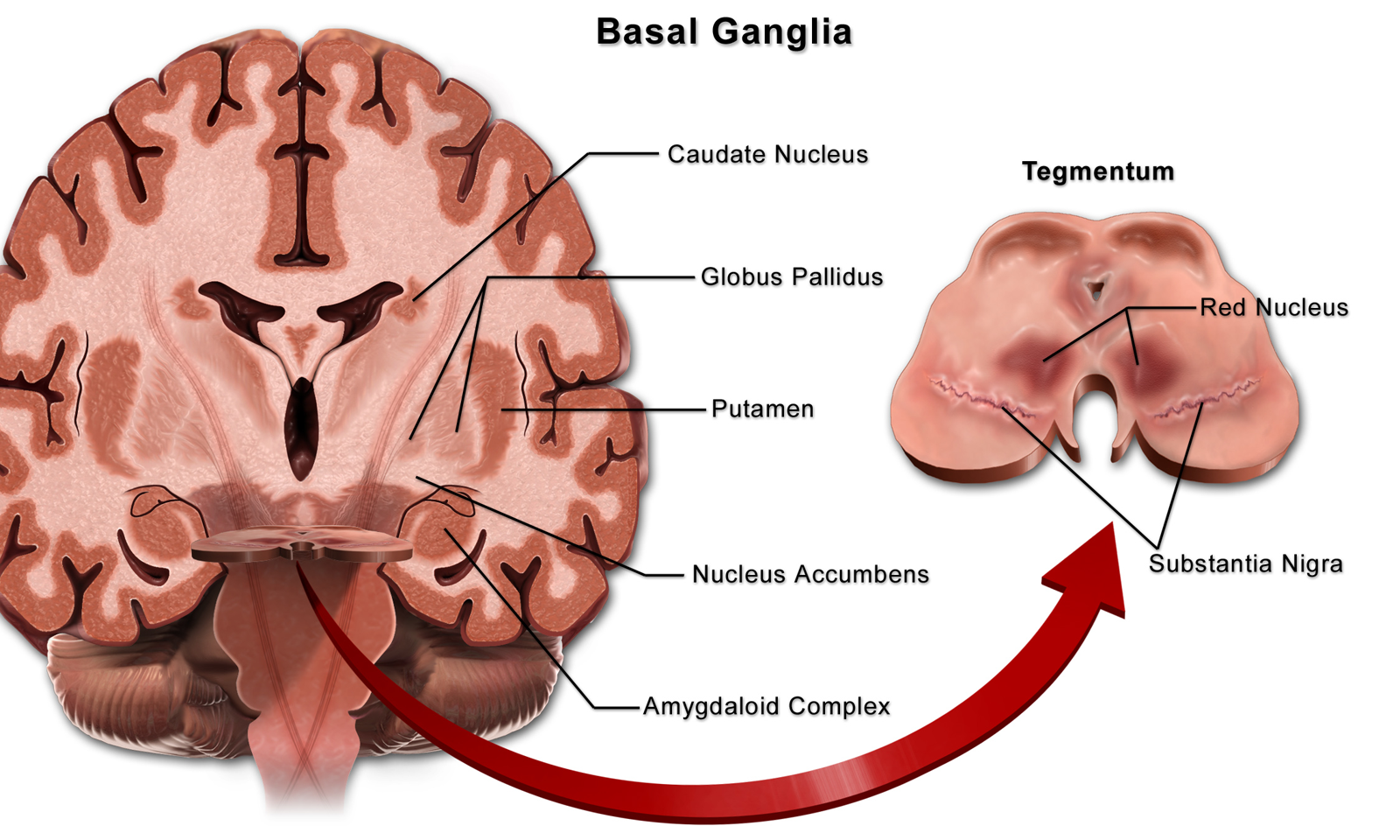
The basal nuclei (blue structures in the diagram) are essential in the control of movement, and regulation of mood and complex behaviors.
An older name for the basal nuclei is basal ganglia. However, we’ve made a big deal of telling you that a “ganglion” is a cluster of nerve cells outside the CNS, and the basal nuclei are definitely part of the CNS, so we’d be hypocritical and confusing to call them “ganglia”, right? That doesn’t stop some neuroscientists from calling it that, but it should.
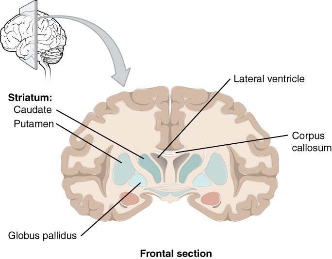 There are three structures that make up the basal nuclei:
- caudate nucleus (Latin: “tail”; remember “caudal” as an anatomical direction means “toward the tail”);
- putamen (Latin: “shell” or “crust”);
- globus pallidus (Latin: “pale globe”).
The basal nuclei receive dopamine from the substantia nigra of the midbrain; when the nigra dies, the lack of dopamine causes the movement disorders of Parkinson Disease. In about half of Parkinson patients, mental disorders also result. Another degenerative disease of the basal nuclei, Huntington Disease, also results in mental disorders. Obsessive-compulsive patients show abnormalities in the caudate nucleus.
Media Attributions
- U11-064 Human Brain Lateral View © Beal, John A. PhD is licensed under a CC BY (Attribution) license
- U11-055 Neural Crest and Neural Tube © Betts, J. Gordon; Young, Kelly A.; Wise, James A.; Johnson, Eddie; Poe, Brandon; Kruse, Dean H. Korol, Oksana; Johnson, Jody E.; Womble, Mark & DeSaix, Peter adapted by Jim Hutchins is licensed under a CC BY (Attribution) license
- U11-056 Primary and Secondary Brain Vesicles © Betts, J. Gordon; Young, Kelly A.; Wise, James A.; Johnson, Eddie; Poe, Brandon; Kruse, Dean H. Korol, Oksana; Johnson, Jody E.; Womble, Mark & DeSaix, Peter adapted by Jim Hutchins is licensed under a CC BY (Attribution) license
- U11-057 Table of Structures © Hutchins, Jim is licensed under a CC BY-SA (Attribution ShareAlike) license
- U11-058 Table of Structures 2 © Hutchins, Jim is licensed under a CC BY-SA (Attribution ShareAlike) license
- U11-059 Table of Structures 3 © Hutchins, Jim is licensed under a CC BY-SA (Attribution ShareAlike) license
- U11-060 Anatomy of the Brain v2 © Jim Hutchins | BioRender is licensed under a CC BY-NC-ND (Attribution NonCommercial NoDerivatives) license
- U11-060a Corpus Callosum © Hutchins, Jim is licensed under a CC BY-SA (Attribution ShareAlike) license
- U11-061 Lobes of the Brain Labeled © Carter, Henry Vandyke adapted by Jim Hutchins is licensed under a Public Domain license
- U11-062a Frontal Lobe © Carter, Henry Vandyke adapted by Jim Hutchins is licensed under a Public Domain license
- U11-062b Parietal Lobe © Carter, Henry Vandyke adapted by Jim Hutchins is licensed under a Public Domain license
- U11-062c Temporal Lobe © Carter, Henry Vandyke adapted by Jim Hutchins is licensed under a Public Domain license
- U11-062d Occipital Lobe © Carter, Henry Vandyke adapted by Jim Hutchins is licensed under a Public Domain license
- U11-063 Insula Colored Area 3 © Carter, Henry Vandyke adapted by Crookston, Alexa is licensed under a CC BY-SA (Attribution ShareAlike) license
- U11-066 Lobes of the Brain Labeled © Carter, Henry Vandyke adapted by Jim Hutchins is licensed under a Public Domain license
- U11-067 Longitudinal Fissure of Cerebrum © Life Science Databases(LSDB) is licensed under a CC BY-SA (Attribution ShareAlike) license
- U11-068 Central Sulcus 1 © Carter, Henry Vandyke adapted by Jim Hutchins is licensed under a CC BY-SA (Attribution ShareAlike) license
- U11-069 Lateral Sulcus © Carter, Henry Vandyke adapted by Jim Hutchins is licensed under a CC BY-SA (Attribution ShareAlike) license
- U11-070 Precentral Gyrus Superior View © Anatomography is licensed under a CC BY-SA (Attribution ShareAlike) license
- U11-070a Precentral Gyrus Lateral View © Anatomography is licensed under a CC BY-SA (Attribution ShareAlike) license
- U11-070b Precentral Gyrus © Carter, Henry Vandyke adapted by Jim Hutchins is licensed under a CC BY-SA (Attribution ShareAlike) license
- U11-071 Postcentral Gyrus Superior View © Anatomography is licensed under a CC BY-SA (Attribution ShareAlike) license
- U11-071a Postcentral Gyrus Lateral View © Anatomography is licensed under a CC BY-SA (Attribution ShareAlike) license
- U11-072 Postcentral Gyrus © Carter, Henry Vandyke adapted by Jim Hutchins is licensed under a CC BY-SA (Attribution ShareAlike) license
- U11-073 Midsagittal Human Brain Dissected Parieto-occipital Sulcus Highlighted © Gumus, Hikmet adapted by Jim Hutchins is licensed under a CC BY-SA (Attribution ShareAlike) license
- U11-074 Parieto-occipital Sulcus © Hutchins, Jim is licensed under a CC BY-SA (Attribution ShareAlike) license
- U11-060 Anatomy of the Brain © Hutchins, Jim is licensed under a CC BY-NC-ND (Attribution NonCommercial NoDerivatives) license
- U11-076 Broadmann Map © Betts, J. Gordon; Young, Kelly A.; Wise, James A.; Johnson, Eddie; Poe, Brandon; Kruse, Dean H. Korol, Oksana; Johnson, Jody E.; Womble, Mark & DeSaix, Peter is licensed under a CC BY (Attribution) license
- U11-076a Table of Brodmann Areas © Hutchins, Jim and Bizzell, Lizz is licensed under a CC BY-SA (Attribution ShareAlike) license
- U11-077 Brodmann © Brodmann, Korbinian adapted by Jim Hutchins is licensed under a Public Domain license
- U11-078 Cortical Map 130 Areas © Glasser, Matthew Ph.D., and Van Essen, David Ph.D. adapted by Jim Hutchins is licensed under a CC BY-SA (Attribution ShareAlike) license
- U11-079 Primary Somatosensory Cortex Lateral View with Homunculus © Neuroanatomy, Somatosensory Cortex is licensed under a Public Domain license
- U11-080 Motor Homunculus © ralf@ark.in-berlin.de adapted by Jim Hutchins is licensed under a Public Domain license
- U11-082 Posterior Parietal Lobe © Paskari is licensed under a CC BY-SA (Attribution ShareAlike) license
- U11-084 Auditory Cortex © Carter, Henry Vandyke adapted by Jim Hutchins is licensed under a CC BY-SA (Attribution ShareAlike) license
- U11-081 Brodmann Area 4 Lateral © Anatomography is licensed under a CC BY-SA (Attribution ShareAlike) license
- U11-083 Brain Sensory Motor © BruceBlaus is licensed under a CC BY-SA (Attribution ShareAlike) license
- U11 085a Prefrontal Cortex © Hutchins, Jim is licensed under a CC BY-NC-ND (Attribution NonCommercial NoDerivatives) license
- U11-086 Phineas Gage © Unknown is licensed under a Public Domain license
- U11-087 Gage Skull © Harlow, John M. M.D. is licensed under a Public Domain license
- U11-088 Basal Ganglia © BruceBlaus is licensed under a CC BY (Attribution) license
- U11-089 Basal Nuclei © Betts, J. Gordon; Young, Kelly A.; Wise, James A.; Johnson, Eddie; Poe, Brandon; Kruse, Dean H. Korol, Oksana; Johnson, Jody E.; Womble, Mark & DeSaix, Peter is licensed under a CC BY (Attribution) license
