Functions of the Circulatory System and Anatomy of the Heart
Objective 1
Identify: structures of the mediastinum, chambers and valves of the heart, and the great vessels of the heart. Describe the layers of the heart and pericardium. Summarize the histology of the heart wall.
In Unit 15, we began our study of the cardiovascular system with an examination of the fluids carried in the cardiovascular system. Now we continue by looking at the pumps and channels that move that fluid around. These pumps and channels deliver blood to the respiratory system and exchange waste carbon dioxide for essential oxygen. This process, termed internal respiration, is carried out in the blood traveling through the pulmonary circuit.
Nutrients are absorbed into the bloodstream in the digestive organs, especially the small intestines, and processed in the liver. The liver will add glucose and many different kinds of proteins to the deoxygenated blood carried to it by the hepatic portal system. From there, blood returns to the heart and is pumped to the lungs. Once oxygen is picked up in the air sacs of the lungs, the nutrients and oxygen are taken to the tissues and delivered by the capillaries.
The capillaries also pick up hydrogen ions (H+), carbon dioxide (CO2), urea, and other wastes. These wastes are removed by the lungs (H+ and CO2) and by the kidneys (H+, urea, and many other waste chemicals). Thus, the lungs and kidneys together regulate body pH (i.e. H+ concentration), a concept that we’ll approach in detail in other courses. Remember from Unit 2 that pH is regulated by the carbonic acid–bicarbonate buffer system, so in some respects both H+, CO2, and bicarbonate ion (HCO3–) are interchangeable forms of acid, acid, and base, respectively. We will see more about these ions and their role in pH control in Units 17 and 19. However, the systems which regulate pH cannot work without effective blood circulation through both the pulmonary and systemic circuits.
Finally, the red and white blood cells which we studied in Unit 15 have to be delivered to the appropriate locations throughout the body. This is also an important function of the circulatory system.
The Mediastinum
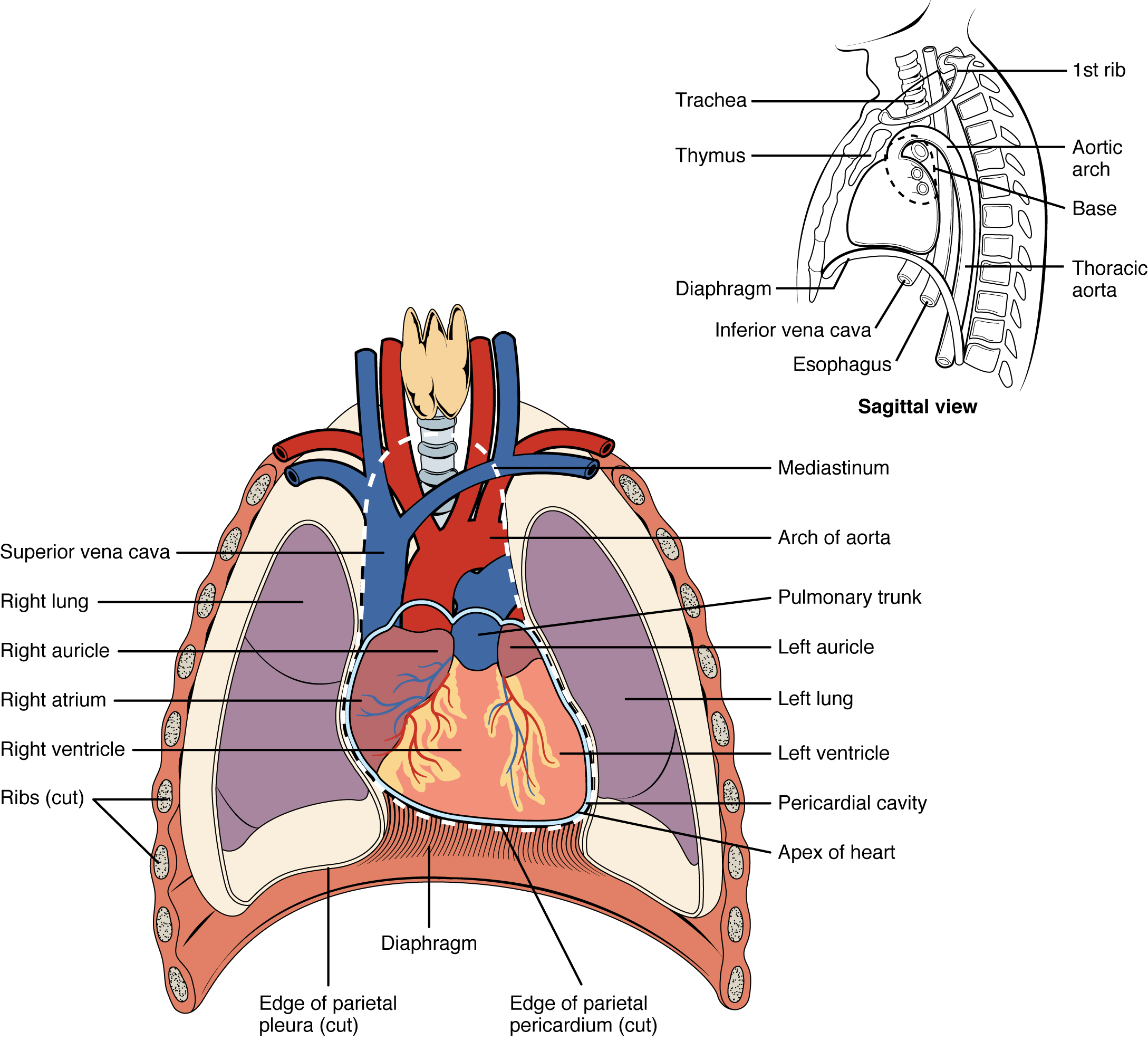
The thorax is the part of the body between the neck and belly. It is defined by the body wall laterally, dorsally, and ventrally. The neck forms the superior border of the thorax and the diaphragm muscle is the inferior border.
The ribs and vertebrae form the bony cage in which the thoracic organs reside.
Most of the volume of the thorax is occupied by the lungs (Unit 17). Everything that is not the lungs is a region called the mediastinum, Latin for “standing in the middle”. Everything that is in the thorax that is not the lungs is the mediastinum. The heart, the great vessels of the heart, parts of the esophagus and trachea, and the thymus all reside within the mediastinum.
Overview of Heart Anatomy
The heart lies almost centrally. A bit more of the heart is on the left side of the midline than on the right. There are great vessels which carry blood into and out of the heart (“great” in this context means “large”, not “wonderful”, although these are indeed useful to have).
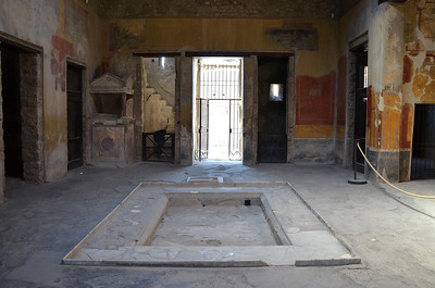
There are two chambers that receive blood called the atria; the atrium of a Roman house was a room with no ceilings and a pool to collect rainwater where the Romans entertained guests.
There are two pumping chambers called the ventricles (Latin: “little belly”).
The Great Vessels
 Veins are vessels which bring blood back to the heart.
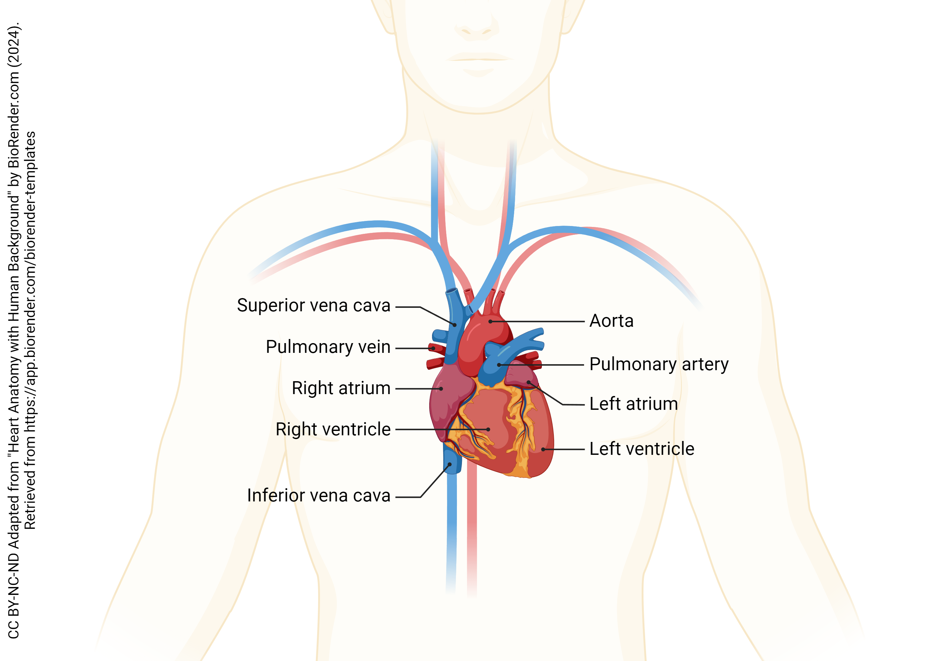
The two largest veins in the human body are the inferior vena cava, which brings blood to the heart from the systemic circulation of the lower body (lower extremity and trunk), and the superior vena cava, which brings blood to the heart from the systemic circulation of the upper body (upper extremity, head, and neck).
Four large veins, the pulmonary veins, bring oxygenated blood from the lungs to the heart. Note that the color of vessels in a diagram indicates the oxygenation status of the blood carried in them and not whether they are veins or arteries. Also, note that deoxygenated blood is not truly blue but a very dark red color.
Arteries are vessels which carry blood away from the heart.
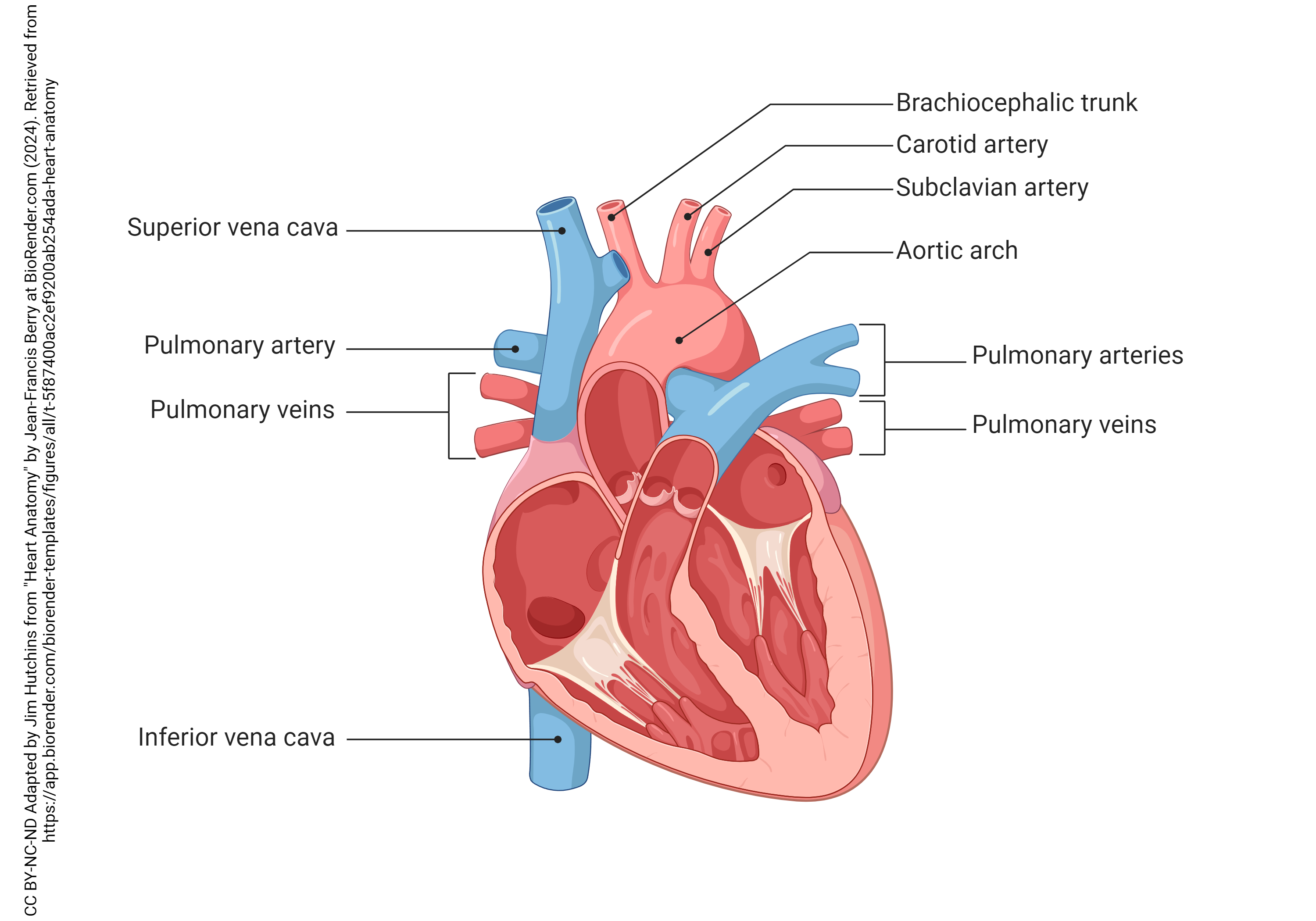
The largest artery in the body is the aorta, a muscular and elastic tube which branches repeatedly to send off major arteries to various parts of the body. Two vessels, the right and left coronary arteries, are the first branches of the aorta (not shown in this diagram but illustrated later).
Another large artery, the pulmonary trunk, divides into right and left pulmonary arteries and then these further subdivide as they supply the lungs with deoxygenated blood.
Chambers of the Heart
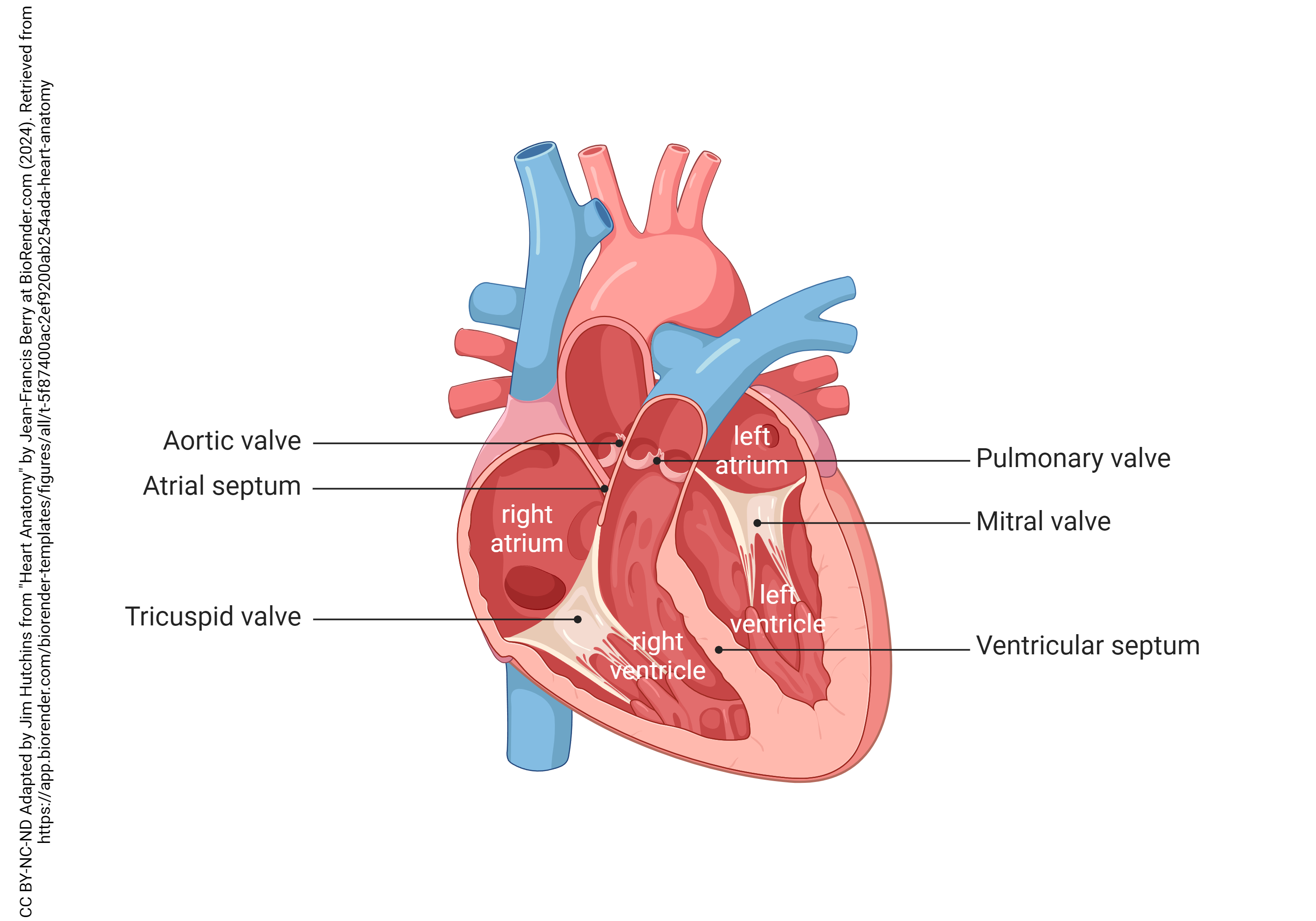
The atria, as mentioned earlier, are the receiving chambers of the heart. The right atrium receives blood from the systemic circuit via the inferior and superior venae cavae. The left atrium receives blood from the lungs via the pulmonary veins.
The ventricles are the pumping chambers of the heart. Accordingly, their muscles are quite strong and the walls of the ventricles are thick. The right ventricle pumps blood to the lungs and the left ventricle pumps blood to the body.
Valves of the Heart
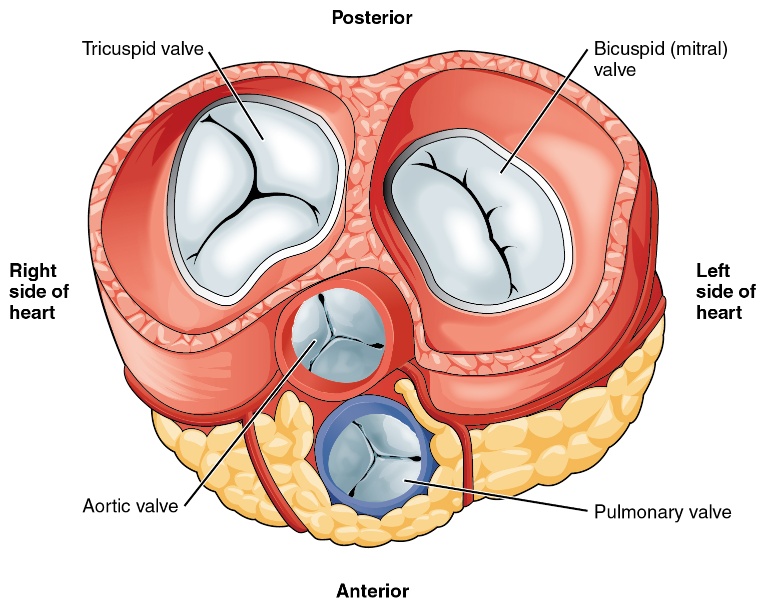 There are two valves connecting each atrium to its same-sided ventricle. Accordingly, these are called atrioventricular (AV) valves.
The right atrium is connected to the right ventricle via the tricuspid valve. The name tricuspid refers to its three leaflets.
The left atrium is connected to the left ventricle via the mitral valve. The name “mitral” refers to its shape, which resembles a bishop’s mitre. The mitral valve is also called the bicuspid valve because it has two leaflets.
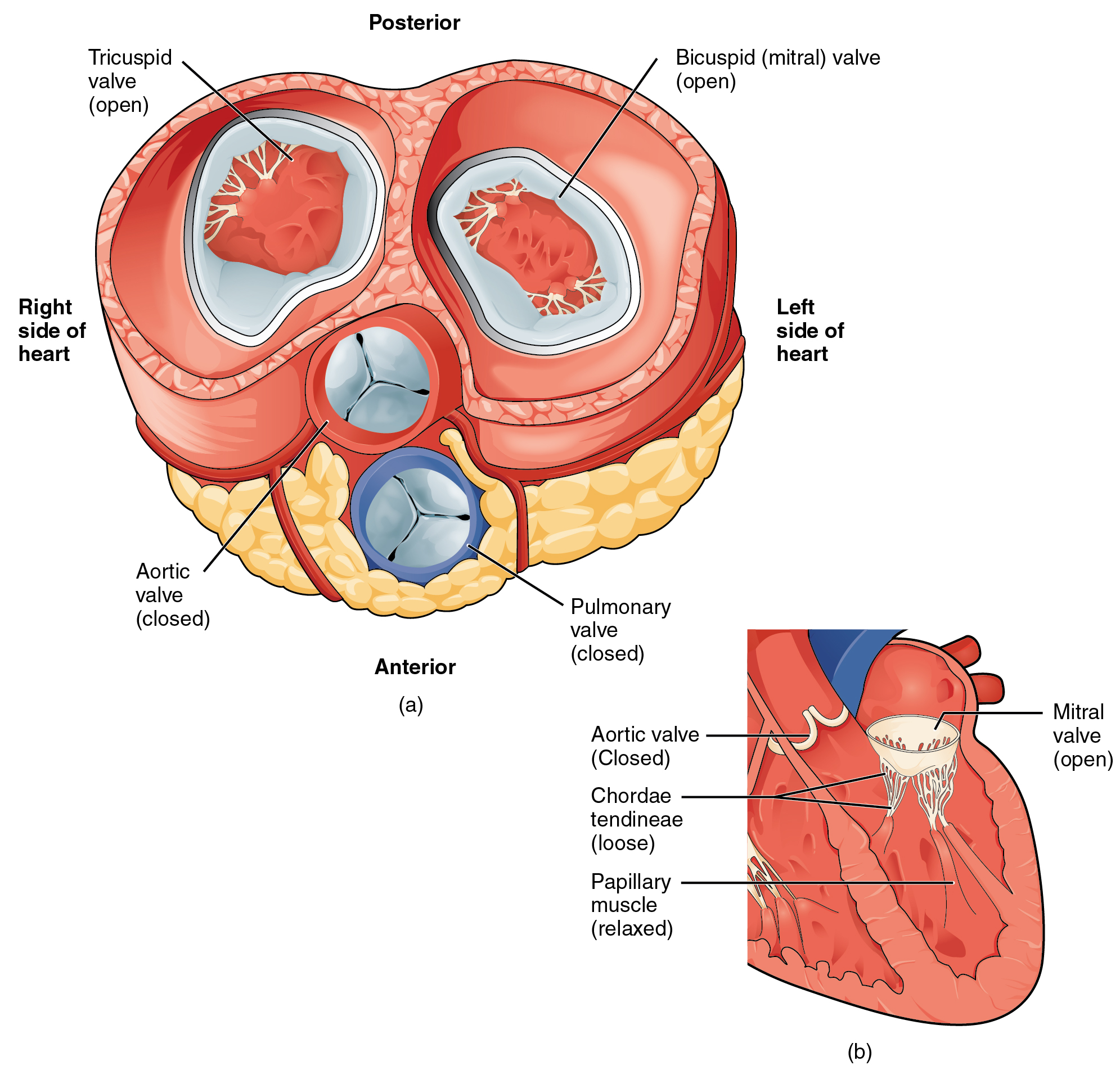
Valves are opened and closed solely by the pressure differences created as different chambers of the heart contract.
For example, the tricuspid and mitral (bicuspid) valves (i.e. the AV valves) are opened by increased pressure in the atrium. They are closed by increased pressure in the ventricles.
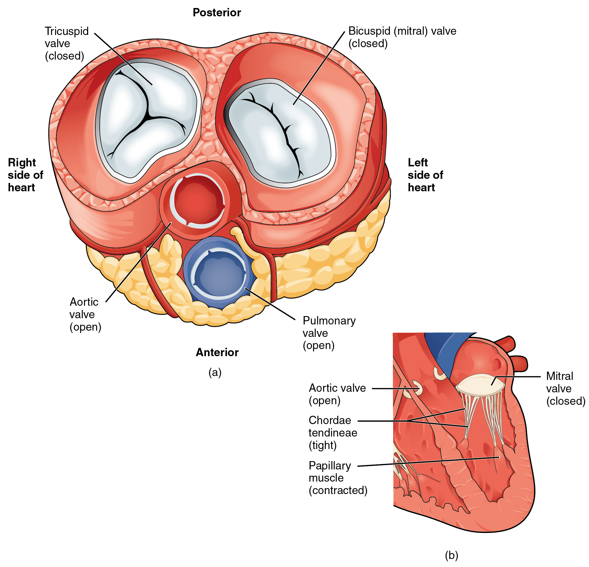 There are two valves which allow blood to leave the ventricles and enter the great vessels. For this reason, these valves are collectively called the outflow valves. Because of their half-moon shape, they are also called semilunar valves. Like the AV valves, the outflow valves operate in pairs. Their names are pretty simple to understand.
The aortic valve opens when the ventricles contract and allows blood to flow from the left ventricle to the aorta.
The pulmonary valve opens when the ventricles contract and allows blood to flow from the right ventricle to the pulmonary trunk.
Layers of the Heart Wall
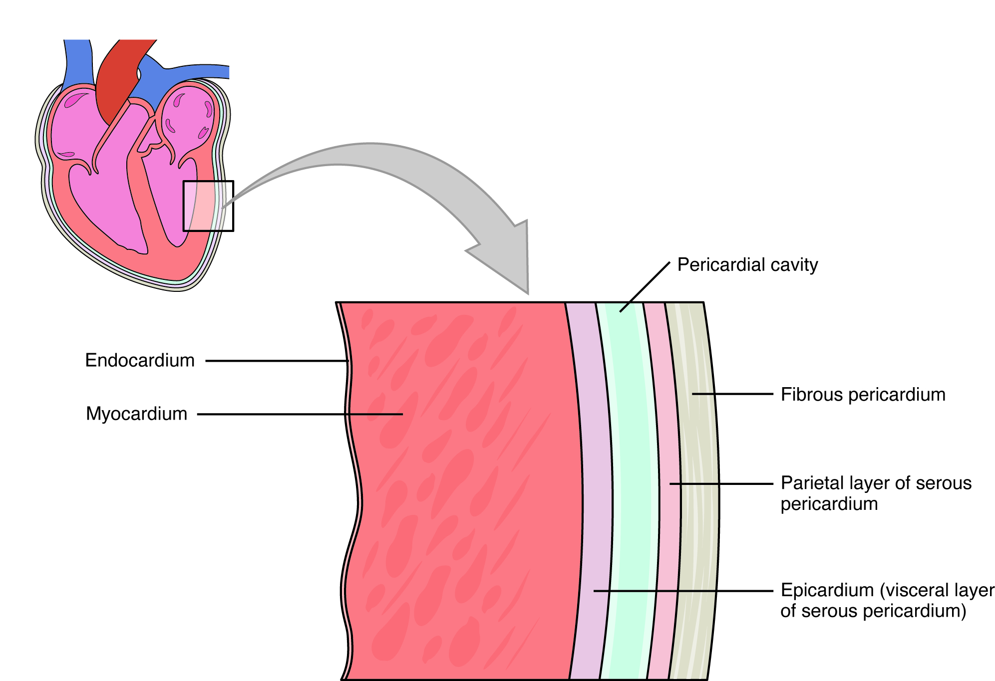 The heart exerts a strong pumping action, and accordingly has a thick muscular layer called the myocardium. It is derived from embryonic blood vessels; like blood vessels, it is lined with an endothelium called the endocardium. The endocardium is a simple squamous epithelium.
Endocardium
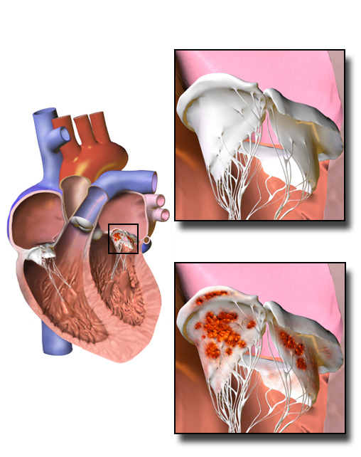 The heart valves are also derived from, and are part of, the endocardium. When the valves or endocardium become infected, this disease is called endocarditis.
It is common to see vegetations in endocarditis. This name refers to visible growths of bacteria or fungi on the surface of the heart valve, which sets up an inflammatory process.
Pericardium
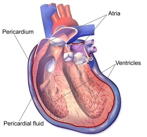 The outside of the heart consists of the pericardium: three layers of tissue and a layer of liquid. The liquid, called pericardial fluid, is contained within a pericardial cavity which is bounded by the two layers of pericardium. Membranes which line organs (including the heart) have two layers: a visceral layer next to the organ itself and a parietal layer closer to the body wall. The visceral layer, closely apposed to the myocardium, is also called the epicardium. The pericardial fluid between the epicardium and the parietal layer of the serous pericardium lubricates the heart and reduces the friction between the layers of the pericardium.
Bounding the entire pericardium is a strong, resilient layer of dense irregular connective tissue called the fibrous pericardium.
Histology of the Myocardium

Cardiac muscle is a kind of striated muscle, but unlike skeletal muscle, it has two important specializations.
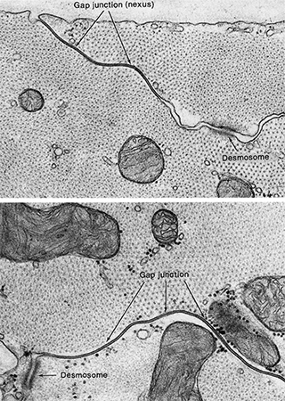 The “spot welds” known as desmosomes (Unit 7) are densely packed in a structure with finger-like extensions of the muscle cell interwoven with each other. While desmosomes are only visible through the electron microscope, these arrays of multiple desmosomes packed densely into the interdigitated extensions of adjacent cardiac muscle cells are called intercalated discs. (The word “intercalated” refers to the Roman practice of inserting extra months into the calendar; similarly, these discs are inserted between each other with desmosomes all over which gives the myocardium tremendous mechanical strength. This keeps the heart muscle from tearing when placed under stress.
Under the electron microscope, desmosomes are visible as threads of cytoskeletal proteins (intermediate filaments) that connect to a dark plate between the cells made of proteins called cadherins (calcium + adherent + –in, protein word ending).
The second specialization of cardiac muscle cells are gap junctions, which allow charged ions to pass rapidly between cardiac muscle cells. These gap junctions (electrical synapses in the nervous system) allow electrical currents to pass easily. In this way, the entire cardiac muscle contracts simultaneously, a property that will be important for the proper functioning of the heart as we’ll see in Objective 3.
Media Attributions
- U16-001 Heart in the Thoracic Cavity © Betts, J. Gordon; Young, Kelly A.; Wise, James A.; Johnson, Eddie; Poe, Brandon; Kruse, Dean H. Korol, Oksana; Johnson, Jody E.; Womble, Mark & DeSaix, Peter is licensed under a CC BY (Attribution) license
- U16-002 Heart Anatomy with Human Background © Hutchins, Jim is licensed under a CC BY-NC-ND (Attribution NonCommercial NoDerivatives) license
- U16-002a Roman Atrium © Raddato, Carole is licensed under a CC BY-SA (Attribution ShareAlike) license
- U16-003 Heart Anatomy Great Vessels © Berry, Jean-Francis and Hutchins, Jim is licensed under a CC BY-NC-ND (Attribution NonCommercial NoDerivatives) license
- U16-004 Heart Anatomy Valves and Chambers © Berry, Jean-Francis and Hutchins, Jim is licensed under a CC BY-NC-ND (Attribution NonCommercial NoDerivatives) license
- U16-05 Valves of the Heart © Betts, J. Gordon; Young, Kelly A.; Wise, James A.; Johnson, Eddie; Poe, Brandon; Kruse, Dean H. Korol, Oksana; Johnson, Jody E.; Womble, Mark & DeSaix, Peter is licensed under a CC BY (Attribution) license
- U16-006 Mitral and Tricuspid Valves © Betts, J. Gordon; Young, Kelly A.; Wise, James A.; Johnson, Eddie; Poe, Brandon; Kruse, Dean H. Korol, Oksana; Johnson, Jody E.; Womble, Mark & DeSaix, Peter is licensed under a CC BY (Attribution) license
- U16-007 Aortic and Pulmonary Valves © Betts, J. Gordon; Young, Kelly A.; Wise, James A.; Johnson, Eddie; Poe, Brandon; Kruse, Dean H. Korol, Oksana; Johnson, Jody E.; Womble, Mark & DeSaix, Peter is licensed under a CC BY (Attribution) license
- U16-008 Heart Wall Layers © Betts, J. Gordon; Young, Kelly A.; Wise, James A.; Johnson, Eddie; Poe, Brandon; Kruse, Dean H. Korol, Oksana; Johnson, Jody E.; Womble, Mark & DeSaix, Peter is licensed under a CC BY (Attribution) license
- U16-008a Endocarditis © BruceBlaus is licensed under a CC BY-SA (Attribution ShareAlike) license
- U16-008b Pericarditis © BruceBlaus is licensed under a CC BY-SA (Attribution ShareAlike) license
- U16-009 Histology of the Heart © Betts, J. Gordon; Young, Kelly A.; Wise, James A.; Johnson, Eddie; Poe, Brandon; Kruse, Dean H. Korol, Oksana; Johnson, Jody E.; Womble, Mark & DeSaix, Peter is licensed under a CC BY (Attribution) license
- U16-009a Desmosomes © Fawcett, Don is licensed under a CC BY-NC-ND (Attribution NonCommercial NoDerivatives) license
