Blood Flow in the Heart | Pulmonary and Systemic Circuits
Objective 2
Describe the blood flow in the heart. Sequence the structures of the pulmonary circuit and of the systemic circuit.
Review of the Chambers and Valves of the Heart
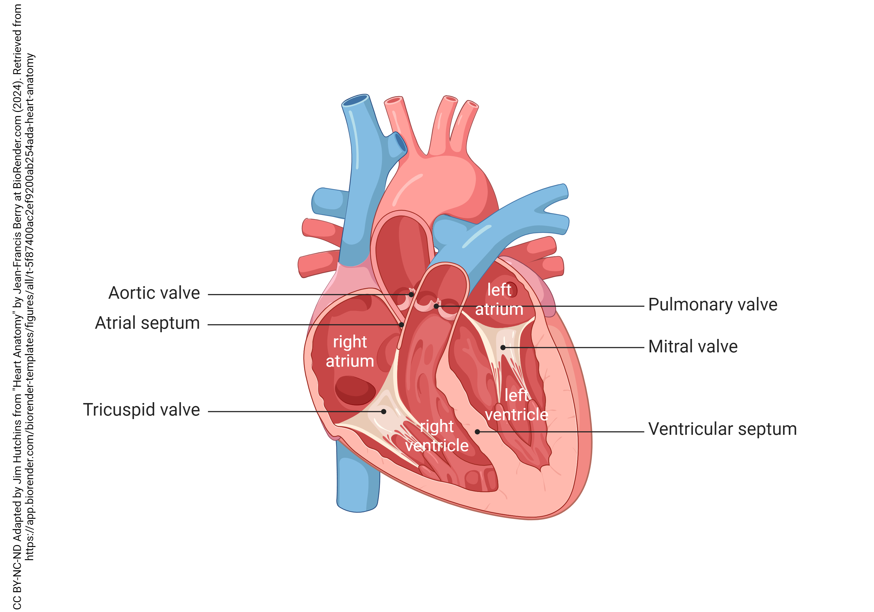
This image, which we discussed previously, shows the chambers and valves of the heart that we will now examine as part of the pulmonary and systemic circuits.
The Ventricles as Pumps
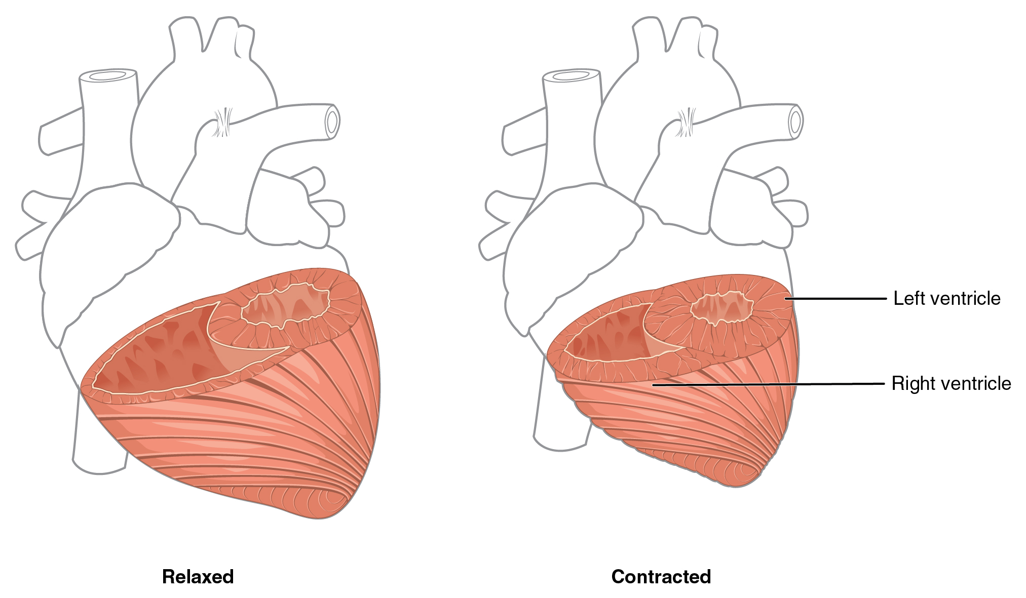 The ventricles are the pumping chambers of the heart. As such, they have the thickest muscular walls. When relaxed, the walls are at their thinnest and the chambers fill with blood. Both ventricles contract at the same time. When they contract, note how the walls thicken as the cardiac muscle cells are squeezed together. Also note how the size of the chamber decreases to about 40% of its previous volume. The (about) 60% of the blood in the ventricles which is squeezed out of the heart is called the ejection fraction and will be the basis for an important measurement of heart function we’ll discuss as part of the cardiac cycle in Objective 4.
The left ventricle has the much harder job of the two, since it must push against the resistance of all the capillaries throughout the body. Accordingly, it has a much thicker muscular wall. The right ventricle is still a substantial pump, but it pushes blood out of the heart and into the capillary beds of the nearby lungs.
Overview of Blood Circulation
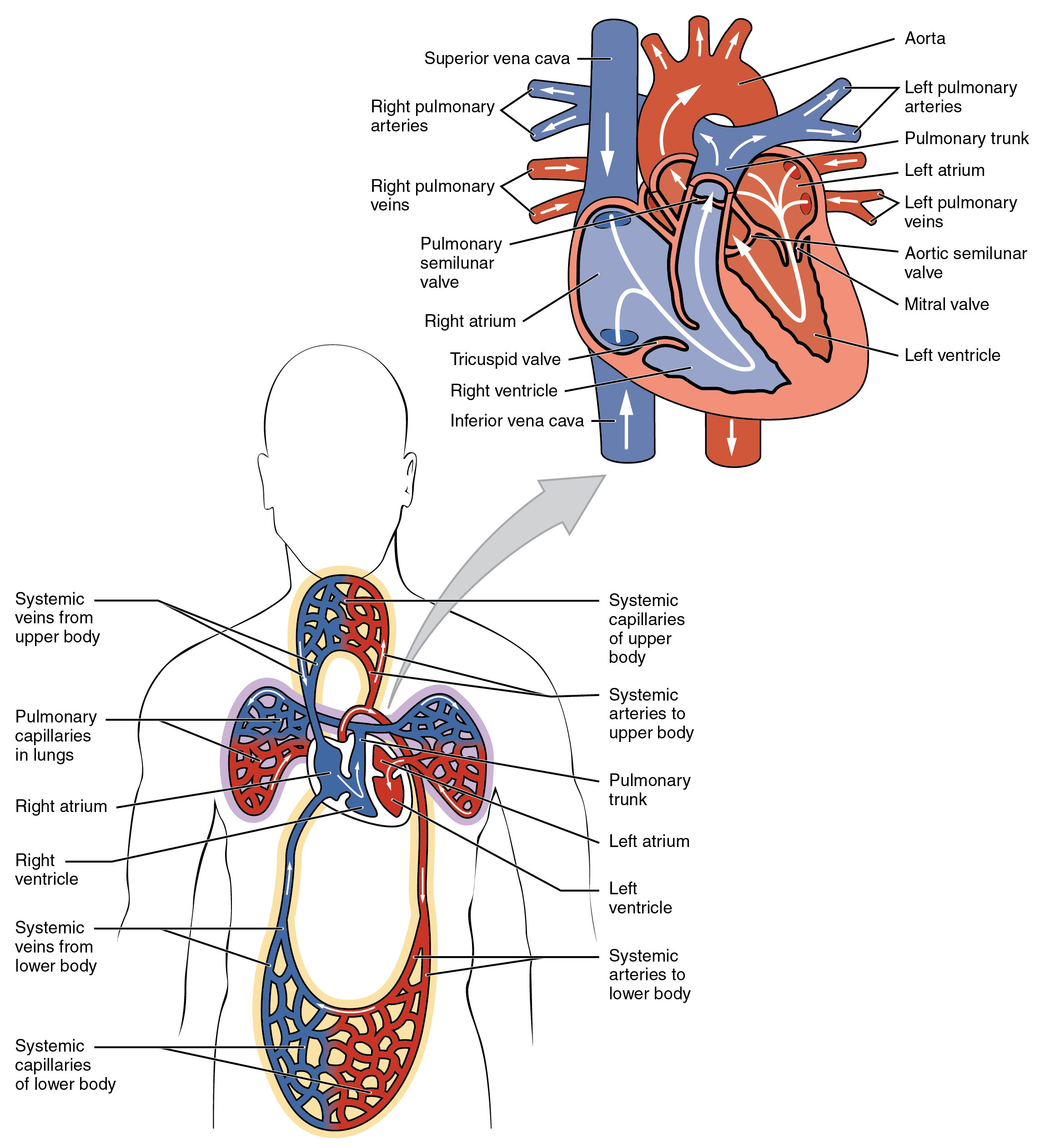 The pulmonary circuit is shown with purple shading in this diagram. The systemic circuit is shown with gold shading.
The Pulmonary Circuit
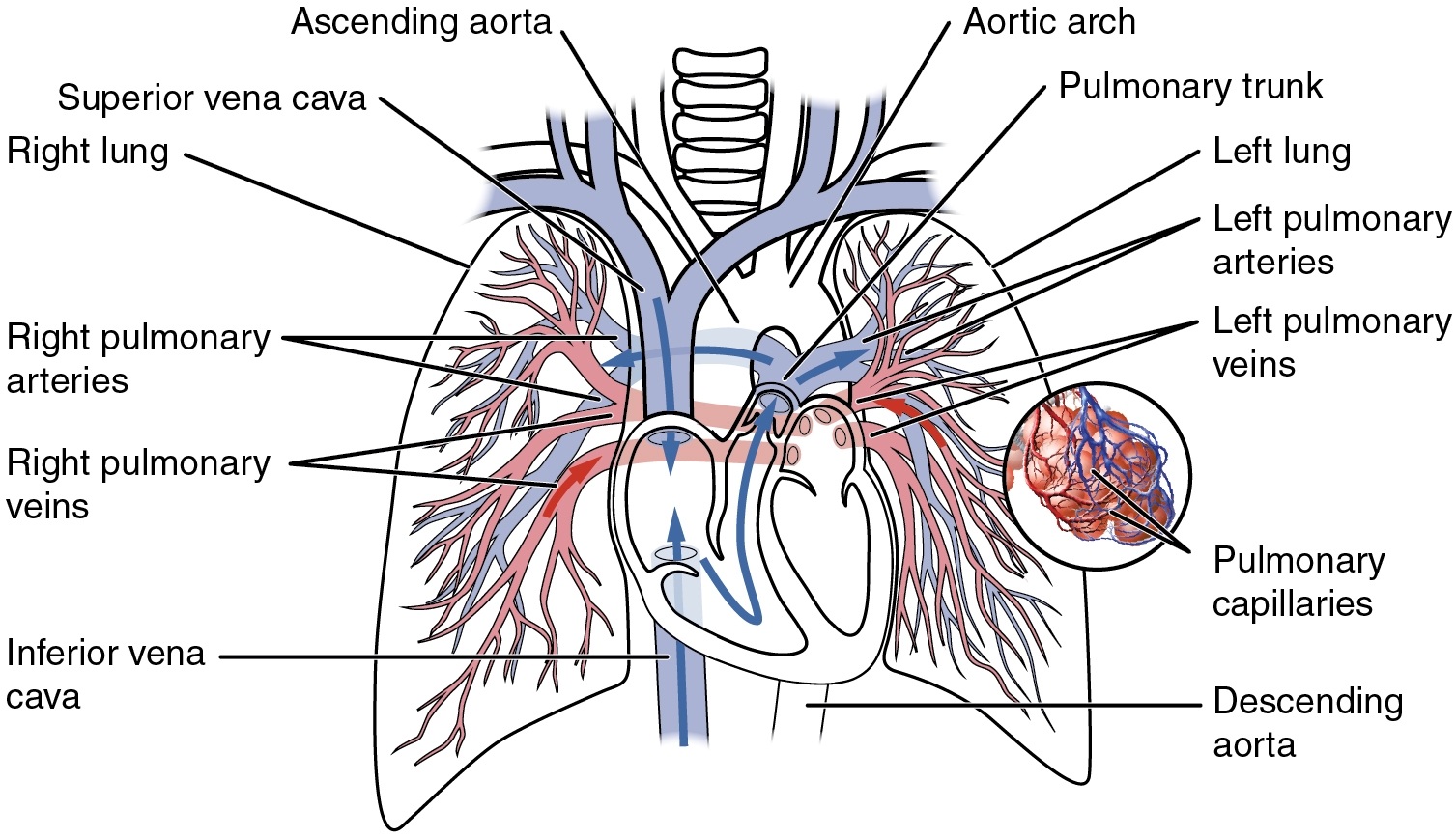 The pulmonary circuit is the mechanism for blood oxygenation. Blood is passed through the capillaries of the lungs where the thin air sacs (alveoli) allow oxygen to diffuse into the capillaries, and allow carbon dioxide to diffuse from the capillaries into the alveoli. As we will see later, the alveoli are quite thin: between the type I alveolar cell, the basement membrane, and the endothelial lining of the capillaries, there is only a distance of 0.5 μm (1/2000 of a millimeter; 1/16 the diameter of a red blood cell).
The pulmonary circuit, beginning at the inferior vena cava and superior vena cava, is:
Venae cavae → right atrium → tricuspid valve → right ventricle → pulmonary valve → pulmonary trunk → right and left pulmonary arteries & their branches → capillary bed of the lung → (left and right) superior and inferior pulmonary veins → left atrium
The Systemic Circuit
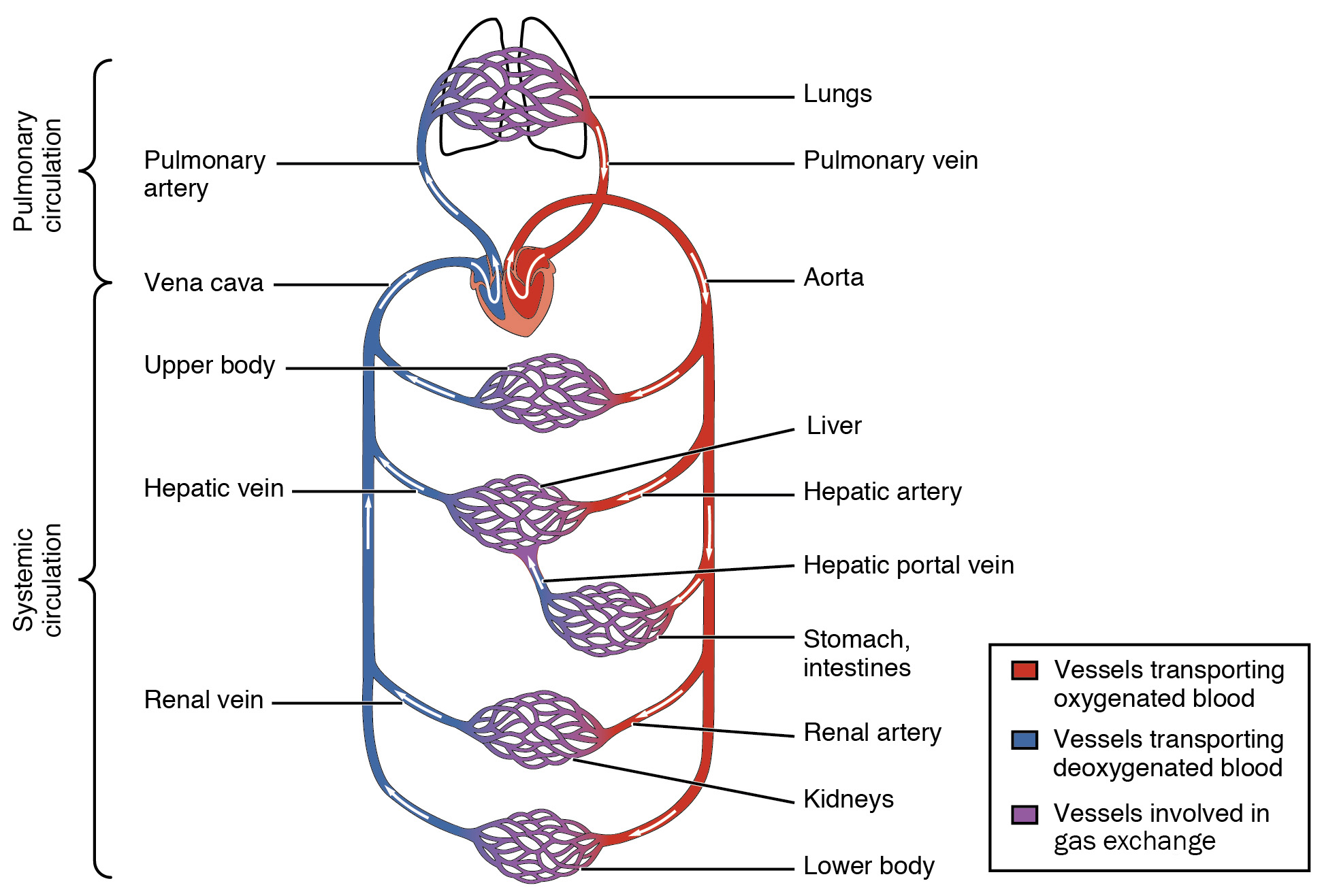 The systemic circuit carries oxygenated, nutrient-rich blood to the body’s capillary network, where it drops off the oxygen, glucose, and other nutrients, and picks up carbon dioxide, hydrogen ions, urea, and other wastes. Depending on the route the blood takes on each circuit, it can be conditioned in various ways on its multiple trips through the systemic circuit. The blood picks up nutrients as it passes through the capillary beds of the stomach and intestines. It drops off carbon dioxide in the lungs. Its pH is adjusted, and its wastes are removed, in the renal capillaries.
(Left and right) superior and inferior pulmonary veins → left atrium → mitral (bicuspid) valve → left ventricle → aortic valve → aorta → systemic arteries → capillary bed of body tissues → systemic veins → superior and inferior venae cavae → right atrium
For the blood passing through the digestive system (stomach and intestines), there is an extra capillary bed, as we will see later and in Unit 18:
(Left and right) superior and inferior pulmonary veins → left atrium → mitral (bicuspid) valve → left ventricle → aortic valve → aorta → systemic arteries → capillary bed of body tissues → hepatic portal vein → capillary bed of liver → inferior vena cava → right atrium
Media Attributions
- U16-004 Heart Anatomy Valves and Chambers © Berry, Jean-Francis and Hutchins, Jim is licensed under a CC BY-NC-ND (Attribution NonCommercial NoDerivatives) license
- U16-011 Contracted and Relaxed States © Betts, J. Gordon; Young, Kelly A.; Wise, James A.; Johnson, Eddie; Poe, Brandon; Kruse, Dean H. Korol, Oksana; Johnson, Jody E.; Womble, Mark & DeSaix, Peter is licensed under a CC BY (Attribution) license
- U16-012 Blood Flow Through the Heart © Betts, J. Gordon; Young, Kelly A.; Wise, James A.; Johnson, Eddie; Poe, Brandon; Kruse, Dean H. Korol, Oksana; Johnson, Jody E.; Womble, Mark & DeSaix, Peter is licensed under a CC BY (Attribution) license
- U16-013 Pulmonary Circuit © Betts, J. Gordon; Young, Kelly A.; Wise, James A.; Johnson, Eddie; Poe, Brandon; Kruse, Dean H. Korol, Oksana; Johnson, Jody E.; Womble, Mark & DeSaix, Peter is licensed under a CC BY (Attribution) license
- U16-014 Blood Flows Through the Systemic Circuit © Betts, J. Gordon; Young, Kelly A.; Wise, James A.; Johnson, Eddie; Poe, Brandon; Kruse, Dean H. Korol, Oksana; Johnson, Jody E.; Womble, Mark & DeSaix, Peter is licensed under a CC BY (Attribution) license
