Adrenal Gland and Adrenal Hormones
Objective 8
Describe the location, structure, and function of the adrenal glands. Explain the interaction of the adrenal medulla with the sympathetic nervous system and identify hormones released as a response. Identify the functional zones of the adrenal cortex. List the hormones and their functions secreted from each zone: 1) the main mineralocorticoid, aldosterone, and its role in the renin-angiotensin-aldosterone system (RAAS), 2) the main glucocorticoid, cortisol, and 3) the androgens (gonadocorticoids). Explain the targets and function of each of these hormones.
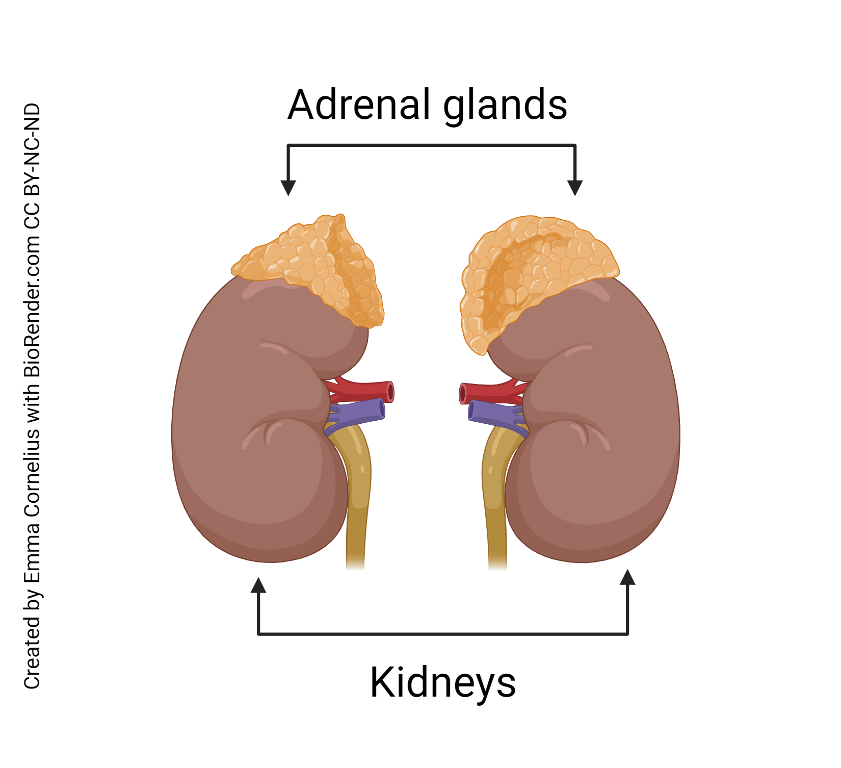 Individuals have a pair of adrenal glands and each one is anatomically superior to one of the kidneys. Each adrenal gland has two distinct regions, the outer adrenal cortex and inner medulla. The cortex comprises 80 to 90% of the gland’s mass.
The adrenal glands are very important to human physiology. They produce steroid hormones that regulate glucose and electrolyte levels. Without sufficient production of these hormones, a person would die within days.
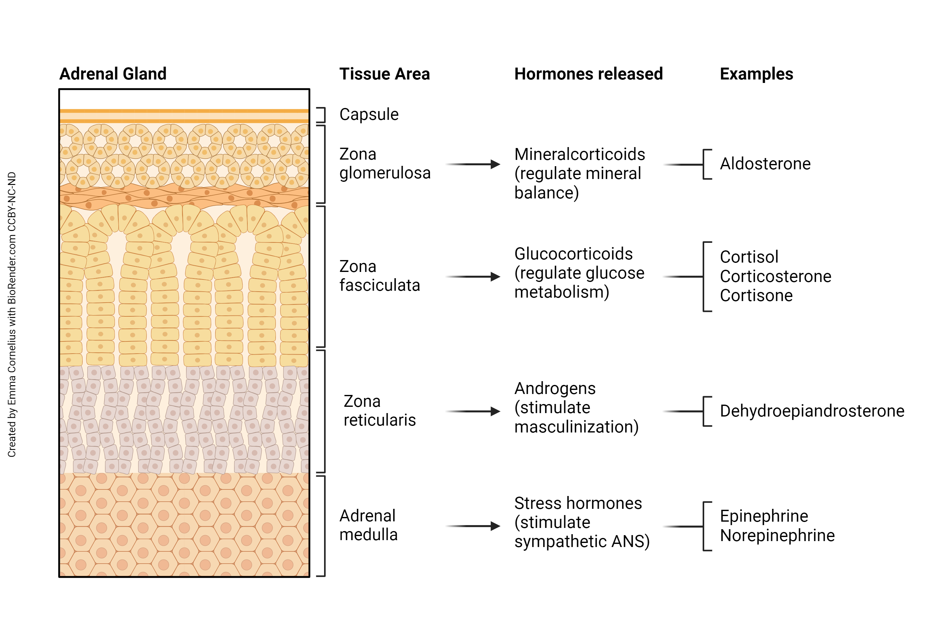
The adrenal medulla is the innermost region of the adrenal glands. The medullae develop as modified sympathetic ganglia of the autonomic nervous system but these do not form post- ganglionic sympathetic neurons. Rather than secreting chemicals as neurotransmitters, secretions released are hormones. The chromaffin cells of the medulla are controlled by sympathetic preganglionic neurons from the CNS. This is why the medullary response is very fast.
The adrenal medullae secrete two hormones, epinephrine and norepinephrine, also known as adrenaline and noradrenaline. We were introduced to these catecholamines in Unit 13. The majority (80%) of the hormone secreted is epinephrine. These hormones are intended to duplicate and prolong a fight-or-flight response from the sympathetic nervous system.
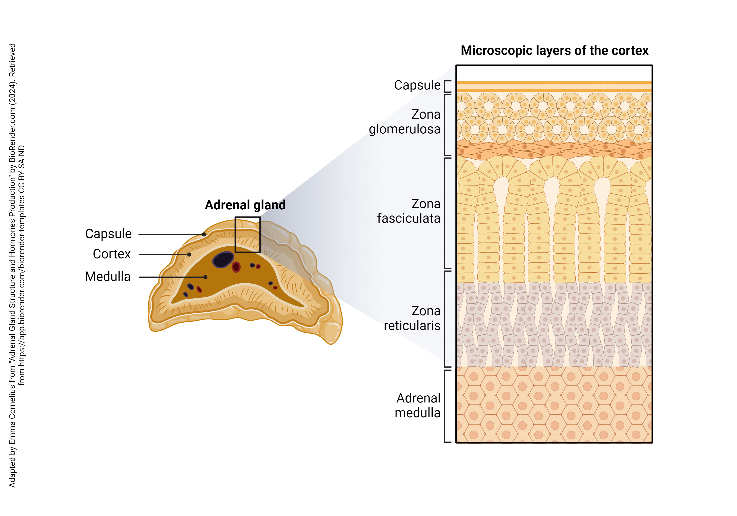
The adrenal cortex consists of three functional zones and produces the “cortical” hormones. These zones, from superficial to deep, are the zona glomerulosa, zona fasciculata, and the zona reticularis. Cells in each of these zones produce and secrete different types of hormones: mineralocorticoids, glucocorticoids, and gonadocorticoids. The names of the different types of hormones provide information about their origin (cortex) and action.
The zona glomerulosa secretes the mineralocorticoids, which function to regulate mineral homeostasis. Aldosterone is the main mineralocorticoid.
The zona fasciculata secretes the glucocorticoids. The primary glucocorticoid is cortisol, and it functions to regulate glucose availability and metabolism.
The zona reticularis secretes small amounts of the gonadocorticoids. These are weak androgens, which are simply masculinizing steroid hormones.
Mineralocorticoids are important in the regulation of salt and water balance. Aldosterone is the main mineralocorticoid. Its principal functions are to increase Na+ and water reabsorption, K+ excretion, and act as a vasoconstrictor. The renin-angiotensin-aldosterone system (RAAS) controls the secretion of aldosterone.
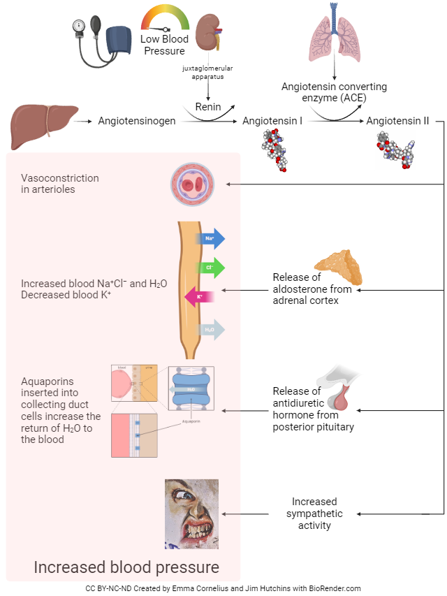
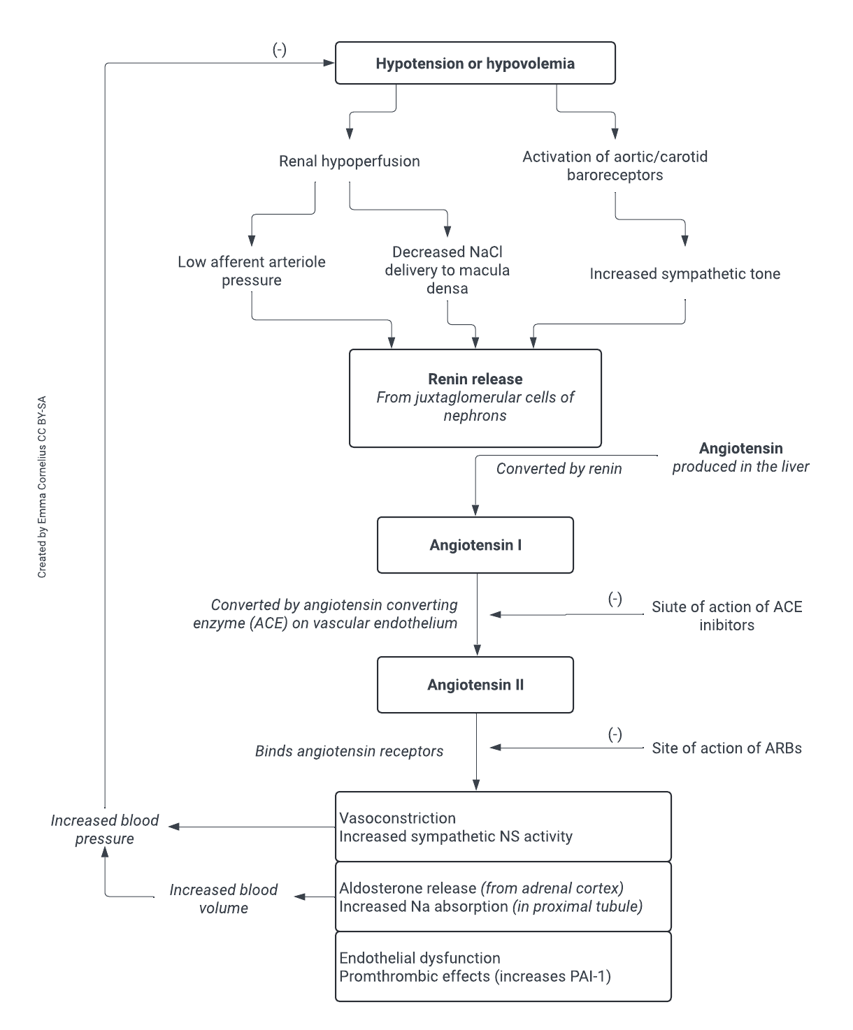
The RAAS can seem very complicated at first. The main stimuli that initiate the RAAS are: low blood sodium, low blood volume, or dehydration. All three of these stimuli are associated with low blood pressure, hence the goal of this system is to raise blood pressure. This is where it all begins:
- The low pressure stimulates a specific group of cells (juxtaglomerular cells) in the kidneys to secrete the enzyme renin.
- Renin circulates in the blood and converts angiotensinogen (a plasma protein produced by the liver) to angiotensin I.
- Angiotensin I continues to circulate in the blood until it comes into contact with an enzyme in the lungs called angiotensin-converting enzyme (ACE). ACE converts angiotensin I to angiotensin II. Angiotensin II has two main actions to increase blood pressure:
- stimulates the contraction of smooth muscle in the walls of arterioles
(vasoconstriction), - stimulates the release of aldosterone from the adrenal cortex.
- stimulates the contraction of smooth muscle in the walls of arterioles
- Aldosterone circulates to the kidneys. Aldosterone increases the reabsorption of Na+ and water, so less is lost in the urine, and stimulates the kidneys to excrete K+ and H+. The increased water reabsorption results in increased blood volume and in turn, increased blood pressure.
Clinical Connection
Many popular medications used to control blood pressure interfere with the RAAS system. One class of drugs, angiotensin-converting enzyme (ACE) inhibitors, block angiotensin converting enzyme from converting angiotensin I to its active form, angiotensin II. Another class of drugs, angiotensin receptor blockers (ARBS) act to block receptors for angiotensin II.
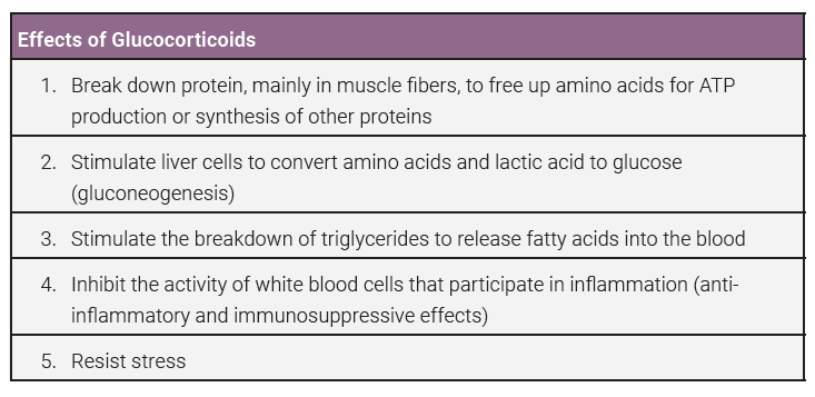
The glucocorticoids regulate glucose metabolism and resistance to stress. Cortisol is the main glucocorticoid. Cortisol wants to make sure there is plenty of glucose for core essential organs that are necessary under stress. For example, if you’re running away from an escaped lion, you’ll need plenty of glucose to supply energy to your heart, lungs, and skeletal muscle. If there isn’t enough available glucose, cortisol will get it from another source, such as protein and lipids. This is called gluconeogenesis.
 Clinical Connection
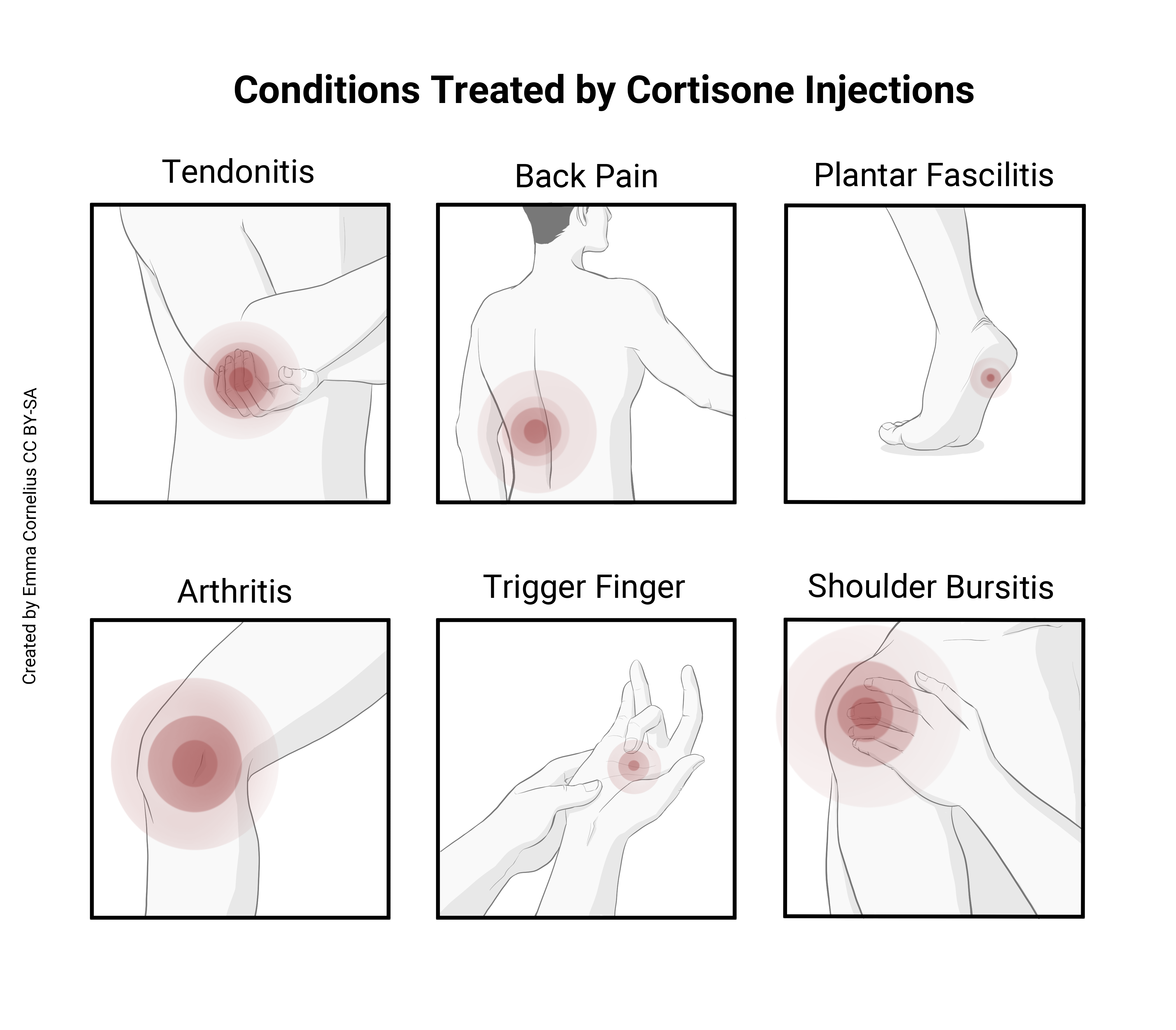
Have you ever had a cortisone injection, taken prednisone for inflammation, or used a hydrocortisone cream for a rash? These are all drugs containing glucocorticoids. One of the effects of glucocorticoids is to suppress the immune system. Cortisone based drugs can be helpful when inflammation causes problems.
Gonadocorticoids are small amounts of steroid sex hormones called androgens. The main androgen secreted by the adrenals is dehydroepiandrosterone (DHEA), which can be converted to testosterone in other tissues. After puberty, the testes in males produce large amounts of testosterone. Adrenal testosterone is negligible in comparison.
In females, adrenal androgens contribute to their libido (sex drive). Because all of the steroid hormones use cholesterol as their backbone, they are similar in structure and can be converted to estrogens in females. After menopause (cessation of ovarian estrogen), the small amount of converted estrogens are likely beneficial.
Media Attributions
- U14-039 Adrenal Gland © Cornelius, Emma is licensed under a CC BY-NC-ND (Attribution NonCommercial NoDerivatives) license
- U14-040 Adrenal Gland Cross Section © Cornelius, Emma is licensed under a CC BY-NC-ND (Attribution NonCommercial NoDerivatives) license
- U14-043 RAAS © Cornelius, Emma and Hutchins, Jim is licensed under a CC BY-NC-ND (Attribution NonCommercial NoDerivatives) license
- U14-044 Flow Chart of the Clinical Effects of RAAS © Cornelius, Emma is licensed under a CC BY-SA (Attribution ShareAlike) license
- U14-045 Effects of Glucocorticoids Table © Newton, Kathy is licensed under a CC BY-SA (Attribution ShareAlike) license
- U14-046 Cortisone Injections © Cornelius, Emma is licensed under a CC BY-SA (Attribution ShareAlike) license
- U14-047 Summary of Adrenal Hormones © Cornelius, Emma is licensed under a CC BY-NC-ND (Attribution NonCommercial NoDerivatives) license
