Female Reproductive Anatomy
Objective 3
Summarize the anatomy of the ovaries, uterine tubes, uterus, vagina, and vulva.
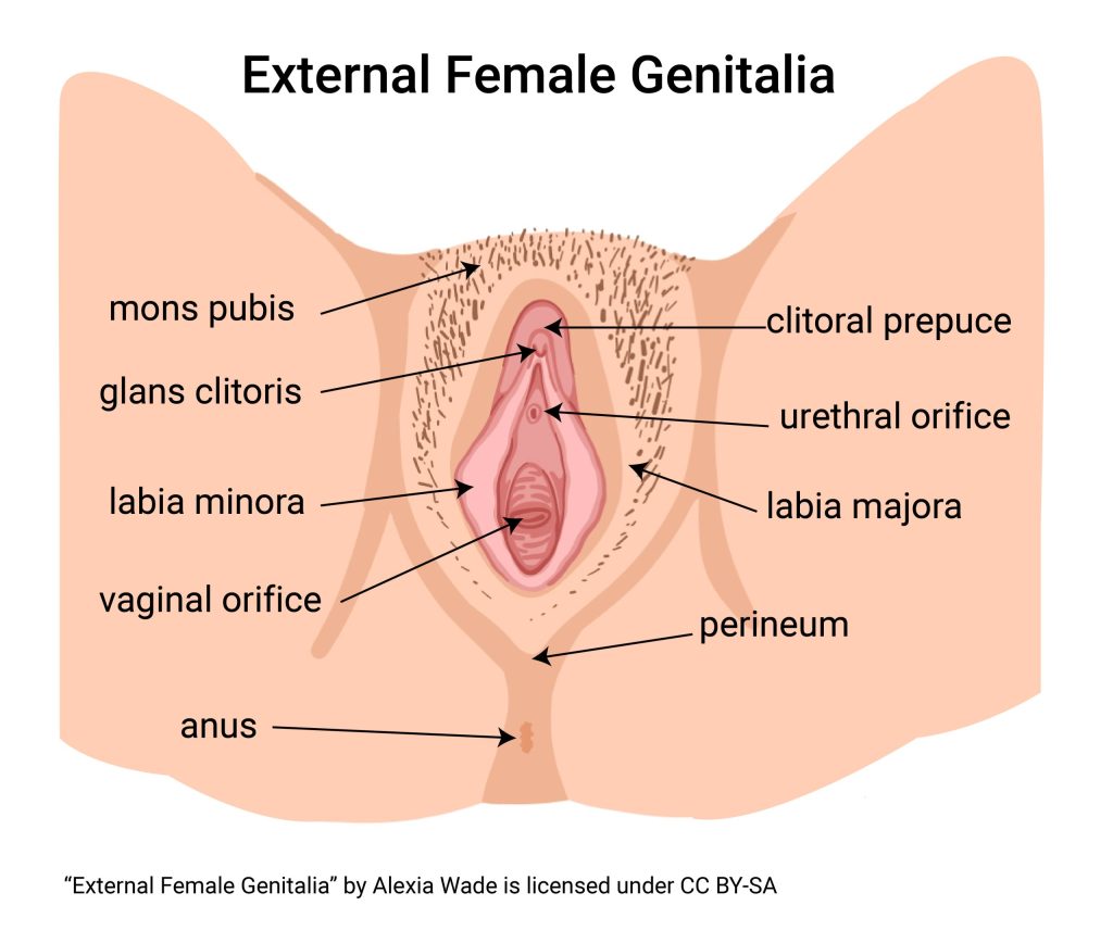
The externally visible parts of the female reproductive system are collectively called the vulva. The female external urethral orifice is part of the vulva, as are the labia minora, labia majora (Latin: “minor lips,” “major lips”), and clitoris. In younger women, a membranous structure called the hymen may partially or completely cover the vaginal orifice.
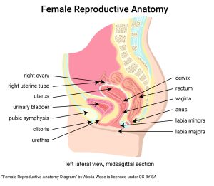
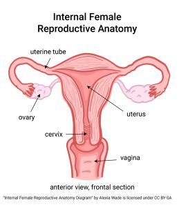
Female gametes are produced in the ovaries. The uterine tubes (Fallopian tubes) convey the gamete from the ovary to the uterus (Latin: “womb”). If fertilization occurs, implantation in the uterine wall follows. If no fertilization occurs or if, for some reason, the embryo is unable to implant (e.g. because of a chromosomal defect), the uterine lining and ovum/embryo are flushed out of the uterus by the process of menstruation.
The cervix (Latin: “neck”) leads out of the uterus into the vagina. Together, during both menstruation and childbirth, they serve as an outlet for menstrual tissue and a channel for childbirth. The vagina is also the location of coitus (sexual intercourse between male and female).
Each of these structures has alternate names:
ovaries = oophor- (Greek ωον- “egg” + -φορα “producing”)
uterine tubes = salpinx (Greek σαλπιγξ, “trumpet”)
uterus = hystero- (Greek υστερα, “womb”) = metra- (Greek μητρα , “womb”)
Clearly, the nomenclature (names of things) is just as complicated as the anatomy. It’s unclear why the Greeks had two names for the womb (hystero- and metra-). The surgical removal of ovaries, uterine tubes, and uterus is called an oophorsalpingohysterectomy or hysterosalpingo-oophorectomy. The first of these is the preferred term.
Media Attributions
- U20-009 External Female Genitalia © Wade, Alexia is licensed under a CC BY-SA (Attribution ShareAlike) license
- U20-008 Female Reproductive Anatomy © Wade, Alexia is licensed under a CC BY-SA (Attribution ShareAlike) license
- U20-010a Internal Female Reproductive Anatomy © Wade, Alexia is licensed under a CC BY-SA (Attribution ShareAlike) license
