Male Reproductive Anatomy
Objective 2
Summarize the anatomy of the scrotum, testes, reproductive ducts, accessory glands, and penis.
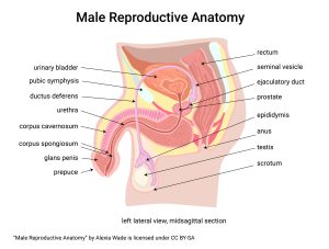
Testes (singular testis), the organs that produce sperm, are enclosed within the scrotum, a sack of skin loosely attached to the body wall. The loose attachment is important because sperm production is most efficient at a temperature about 2-3°C below core body temperature.
The testes require a rich blood supply which arrives and leaves through the spermatic cord, a canal enclosed by connective tissue. The duct which carries sperm from each testis is called the ductus deferens (vas deferens; Latin, ductus “duct” or vas “vessel + deferens “carrying away”) and is also enclosed in the spermatic cord. When the vas deferens is cut, the surgery is called a vasectomy.
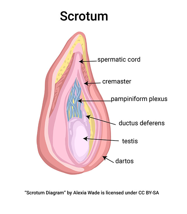
The scrotum has two muscles. The dartos, which surrounds the scrotal sac subcutaneously, will cause wrinkles to appear in the sac when the muscle contracts. The cremaster is a paired muscle (right and left) attached to each testis and is part of the tissue surrounding the spermatic cord. Together, the dartos and cremaster relax to distance the testes from the body or contract to draw the testes closer to the body, thus helping maintain testicular temperature slightly below body temperature.
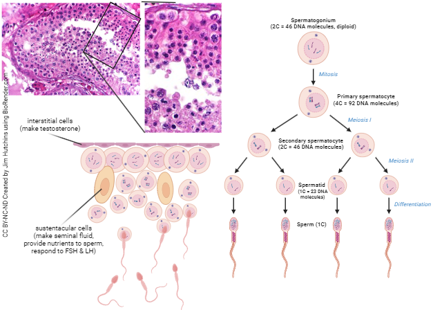
The organs of sperm production are the testes (singular testis). Production of sperm in the testes continues throughout the life of the male, although there are complex changes with age that have just recently been studied. Spermatozoa are made by spermatogenic cells, which are supported by sustentacular cells (Sertoli cells). Interstitial cells (Leydig cells) produce androgens (primarily testosterone), the hormones which support male metabolism and secondary sexual characteristics (characteristics that develop at puberty).
Inside each testis, a series of seminiferous tubules lead to a central collection network (rete testis) which then empties through efferent ducts into the epididymis. The epididymis (Greek επι- “on top of” + -διδυμος “testicles”) is closely associated with the testis inside the scrotal sac.
The spermatozoa produced in the testes need to be delivered to the tip of the penis at ejaculation. Since that is a pretty long journey for a bunch of microscopic haploid flagellated cells, they need a safe liquid medium in which to swim and a sack lunch for nutrition along the way. Guiding them to their destination and taking care of their needs is the job of the ducts and the accessory sex glands.
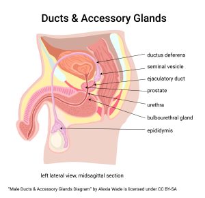 The ducti deferens (paired ducts, one for each testis) carry sperm from the epididymis to the urethra at the prostate gland. The seminal vesicles and bulbourethral glands are also paired, left and right. The prostate gland is a single, midline structure. We will examine these in more detail when we look at their function, but right now we’re just looking at the anatomy.
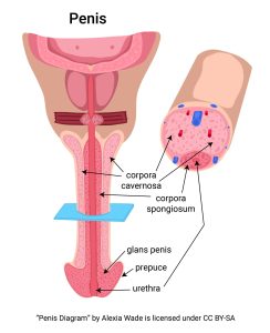
The last structure to consider in male reproductive anatomy is the penis. Erection of the penis is necessary so it can be introduced into the female vagina to deliver sperm at ejaculation. Some are surprised to learn there are no muscles directly involved in penile erection. Rather, erection is all about blood flow—blood is allowed to flow into the penis but outflow is blocked. This venous congestion results in erection. The tissue involved is called erectile tissue and is divided into three chambers:
- the corpus spongiosum (a midline structure that surrounds the urethra and also makes up the bulk of the head of the penis or glans penis), and
- the paired left and
- right corpora cavernosa (singular corpus cavernosum) which make up most of the shaft of the penis.
Uncircumcised men have a prepuce (foreskin) partially covering the glans penis. The prepuce can be retracted to expose the glans penis and then reduced to cover it again. Circumcision is a surgical procedure that removes most of the prepuce.
Media Attributions
- U20-002 Male Reproductive Anatomy © Wade, Alexia is licensed under a CC BY-SA (Attribution ShareAlike) license
- U20-003 Scrotum © Wade, Alexia is licensed under a CC BY-SA (Attribution ShareAlike) license
- U20-004 Spermatogenesis © CoRus13 and BioRender adapted by Jim Hutchins is licensed under a CC BY-NC-ND (Attribution NonCommercial NoDerivatives) license
- U20-005 Testisanimacion © KDS444 is licensed under a CC BY-SA (Attribution ShareAlike) license
- U20-006 Ducts and Accessory Glands Male © Wade, Alexia is licensed under a CC BY-SA (Attribution ShareAlike) license
- U20-007 Penis © Wade, Alexia is licensed under a CC BY-SA (Attribution ShareAlike) license
