Digestion
Objective 9
Define the following phases of digestion: cephalic phase, gastric phase, intestinal phase. Give specific examples of the role of mucous glands, stomach, and small intestines in each phase. Define mechanical digestion. Identify the role of each of the digestive organs in the process of mechanical digestion. Define chemical digestion. Identify the role of each of the digestive organs in the process of chemical digestion. List the enzymes involved in chemical digestion. Define the substrates and products for each enzyme.
Phases of Digestion
Digestion has three phases:
- cephalic phase
- gastric phase
- intestinal phase
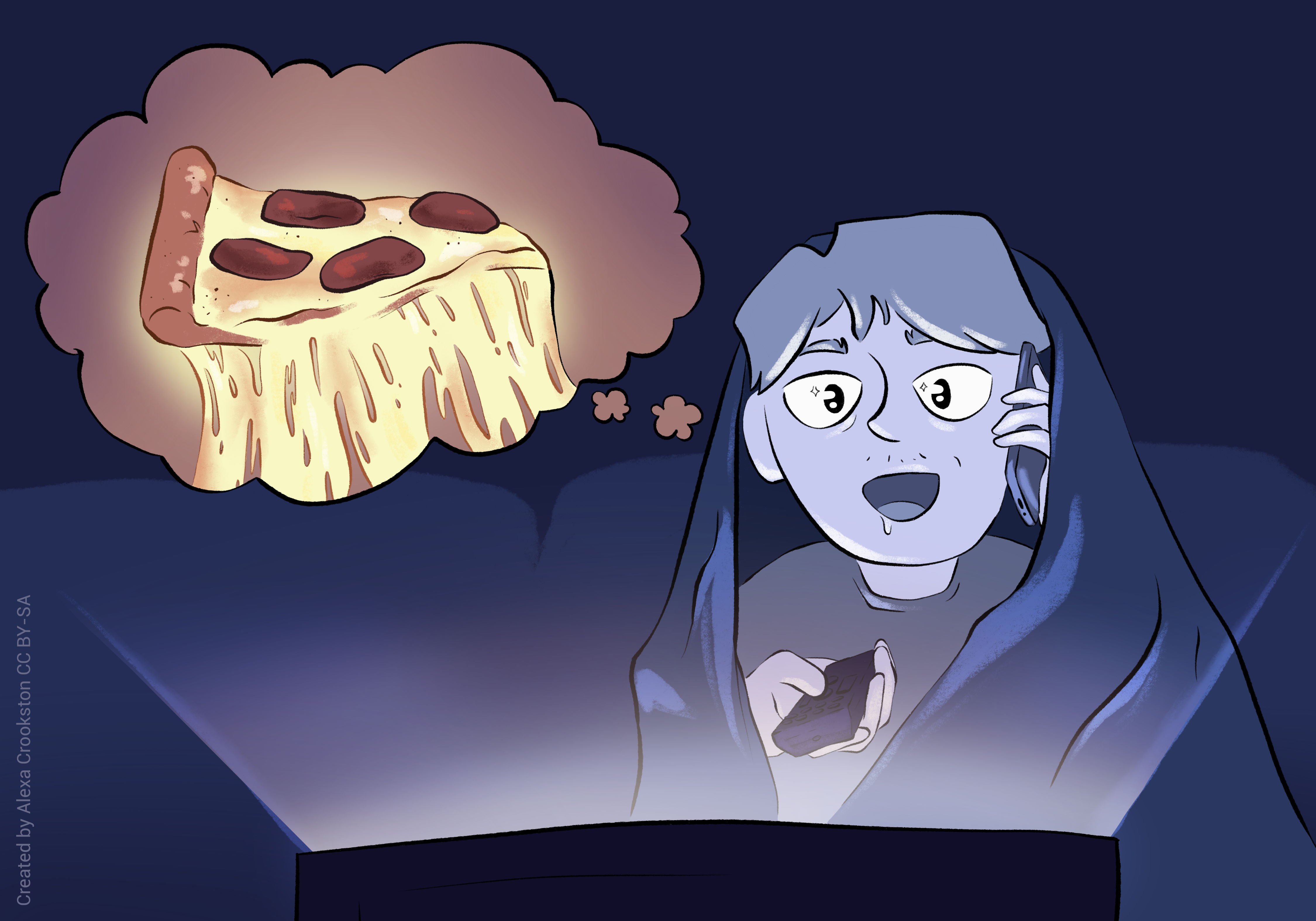
In the cephalic (“head”) phase, the brain is stimulated by the smell, taste, sight, or thought of food. The brain begins to prepare the body to receive a meal. Cranial nerves innervating the salivary glands become active, increasing salivation; the vagus nerve (CN X) releases acetylcholine onto parietal cells, which increases stomach acid secretion. The cephalic phase is manipulated by the wahoos who run pizza ads during major sporting events. You may not even be hungry, but you are salivating and your stomach is full of acid, so you order the stupid pizza.
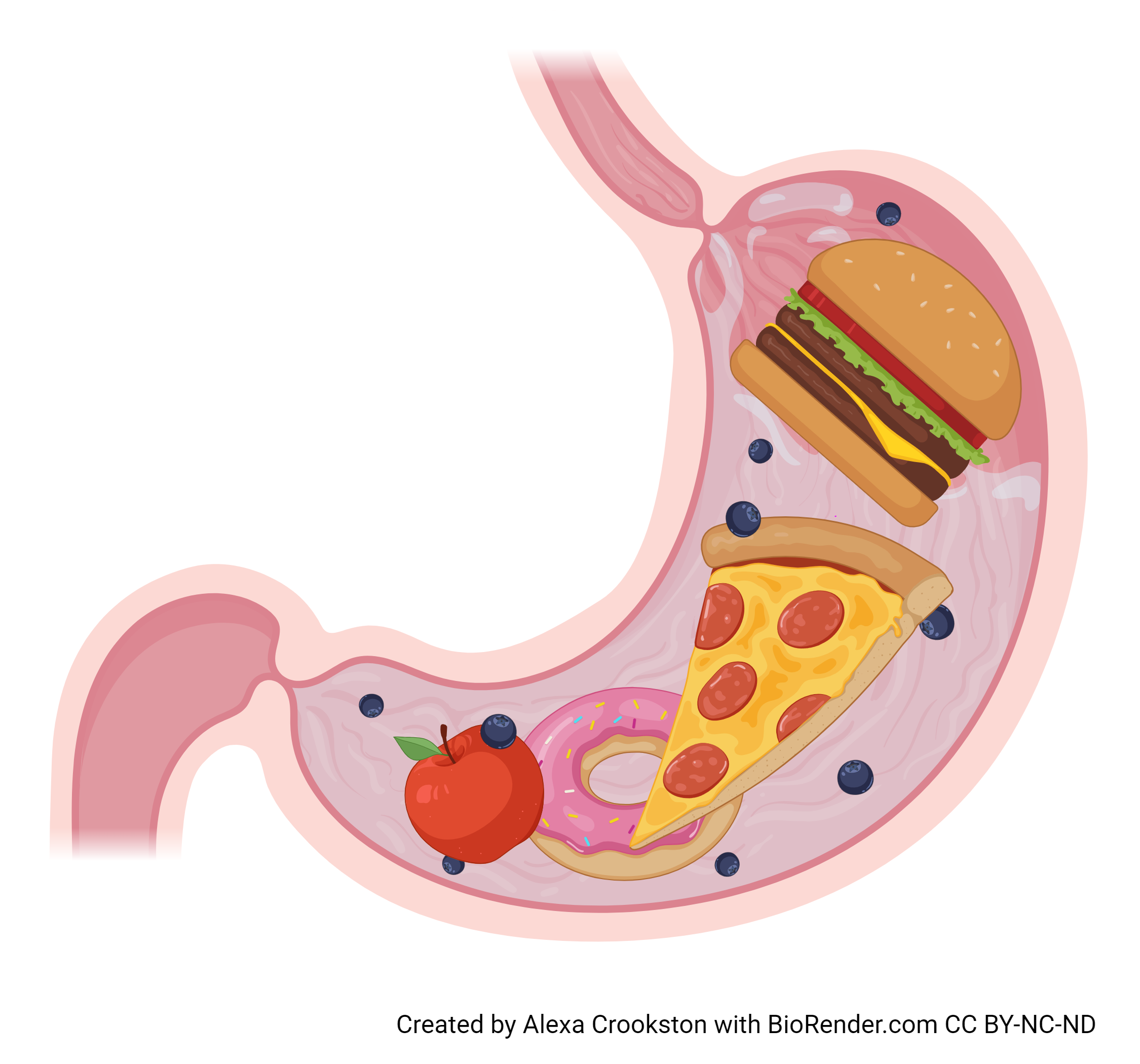
The gastric phase begins when food stretches the stomach wall, or when food buffers the acid in the stomach and stomach pH rises. Gastric motility and HCl secretion are stimulated.
The stomach empties into the duodenum. This phase is regulated by gastrin from G cells of the stomach.
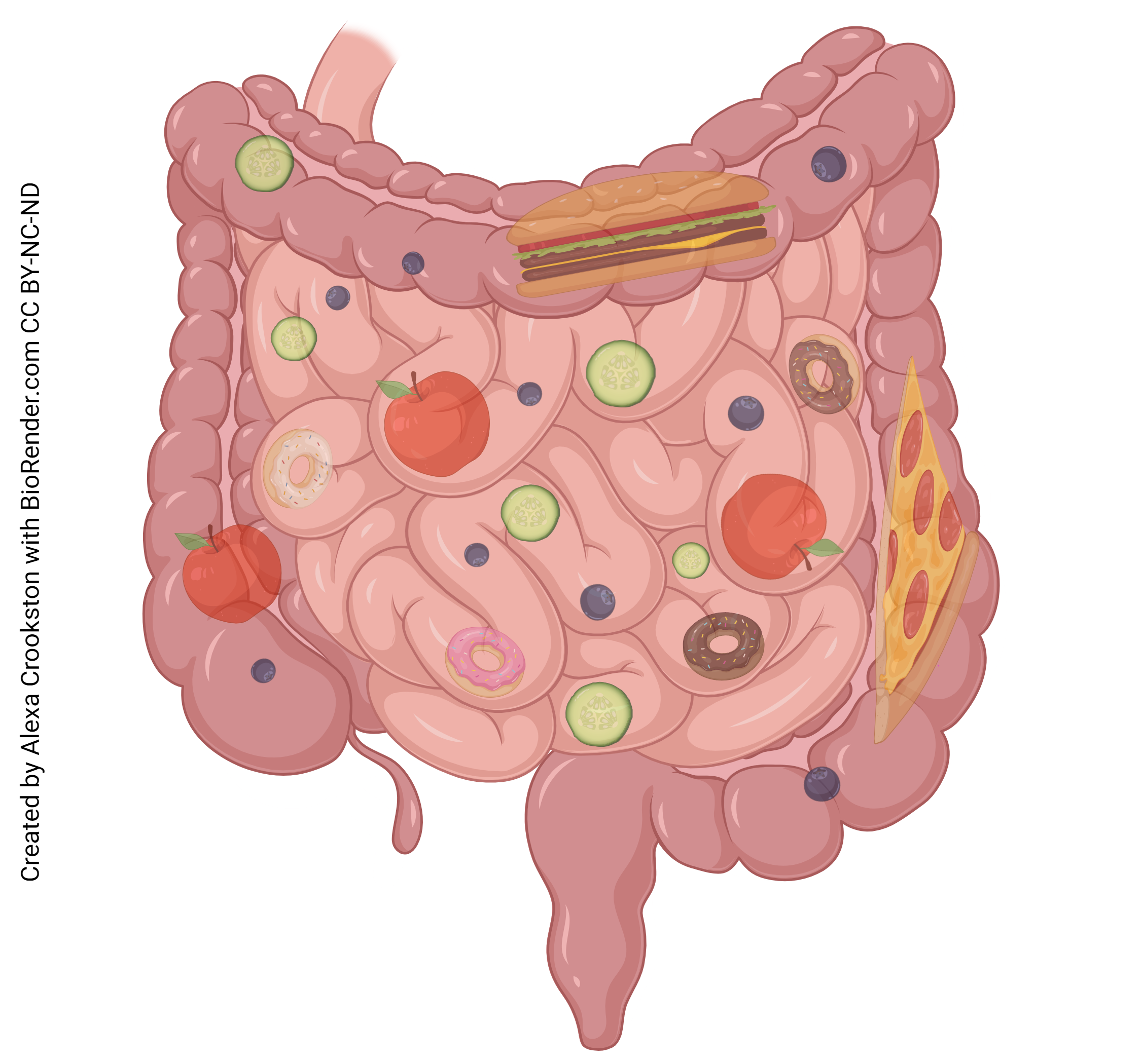 In the intestinal phase, chyme stretches the duodenum. The duodenum signals the medulla, and the medulla in turn shuts down the vagus nerve. This decreases gastric motility and closes the pyloric sphincter. This phase is regulated by the hormone’s cholecystokinin and secretin, both released by the duodenum.
Components of Digestion
Digestion has two components: mechanical and chemical.
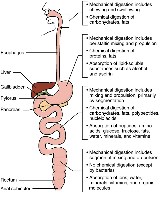
Mechanical digestion includes all the mixing, moving, churning, mashing and ripping forces we’ve discussed.
In the oral cavity, chewing our food thoroughly, (as Mom always told us to) properly begins the process of mechanical digestion.
All along the GI tract, peristalsis aids in mechanical digestion.
Each of the organs along the gut tube has folds and wrinkles to help in the mixing process of mechanical digestion:
- in the stomach, rugae (sing. Latin ruga, “a wrinkle on the face”)
- in the small intestine, plicae circulares
- in the large intestine, haustra (Latin haurere, “to draw up”)
The companion to mechanical digestion is the breakdown of food into nutrients through chemical reactions, or chemical digestion.
Like mechanical digestion, chemical digestion begins as soon as you put food in your mouth. There, the salivary glands secrete enzymes that break down starches (salivary amylase) and fats (lingual lipase).
Further down the GI tract, the stomach secretes HCl to break down all kinds of food. Chief cells secrete pepsinogen which is converted to its active form, the enzyme pepsin, by HCl. Pepsin hydrolyzes protein. Chief cells also secrete gastric lipase (which hydrolyzes fat).
In the duodenum of the small intestine, sodium bicarbonate is excreted by the pancreas and added to the acidic chyme from the pylorus. This buffers the chyme to a pH near 7. Pepsin is inactivated by a pH above 5.
 Enzymes that prefer near-neutral pH are made in the pancreas and released into the duodenum via the hepatopancreatic sphincter. Digestive enzymes are also found along the brush border of the small intestine, and predominate in the small intestine.
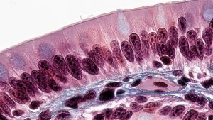
Enzymes in the small intestine which break down various substances found in foods (primarily carbohydrates, proteins, and lipids) are found along a region called the brush border. In the electron microscope, the brush border appears “brushy” because the microvilli are coated with proteins (enzymes), giving them a fuzzy appearance.
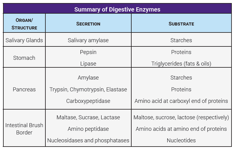
The major enzymes which aid in chemical digestion, and their locations in the GI tract, are listed in the table. You should know a little bit about each one of these enzymes, what it does, and where it is typically found.
Media Attributions
- U18-056 Cephalic Phase © Crookston, Alexa is licensed under a CC BY-SA (Attribution ShareAlike) license
- U18-057 Gastric Phase © Crookston, Alexa is licensed under a CC BY-NC-ND (Attribution NonCommercial NoDerivatives) license
- U18-060a Intestinal Phase © Crooskton, Alexa is licensed under a CC BY-NC-ND (Attribution NonCommercial NoDerivatives) license
- U18-059 Digestion © Betts, J. Gordon; Young, Kelly A.; Wise, James A.; Johnson, Eddie; Poe, Brandon; Kruse, Dean H. Korol, Oksana; Johnson, Jody E.; Womble, Mark & DeSaix, Peter is licensed under a CC BY (Attribution) license
- U18-060b Brush Border © Berkshire Community College Bioscience Image Library is licensed under a CC0 (Creative Commons Zero) license
- U18-061 Table of Digestive Enzymes © Burr, Justin is licensed under a CC BY-SA (Attribution ShareAlike) license

.jpg)