Respiratory System Histology
Objective 4
Describe the histology of the respiratory system.
Remember that function denotes structure. This is true of the tissues of the respiratory tract. Tissues vary throughout the respiratory tract to meet specific functions.
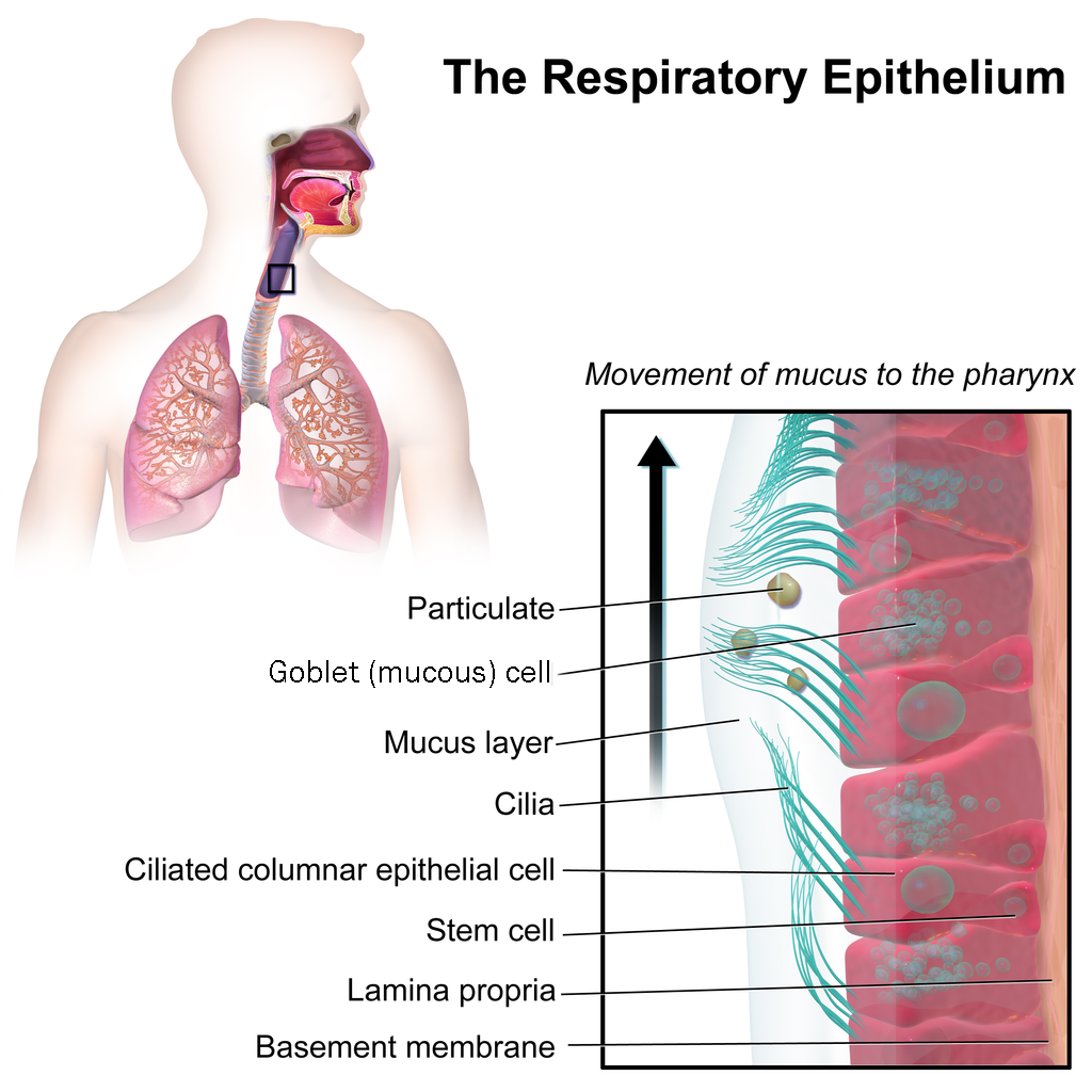
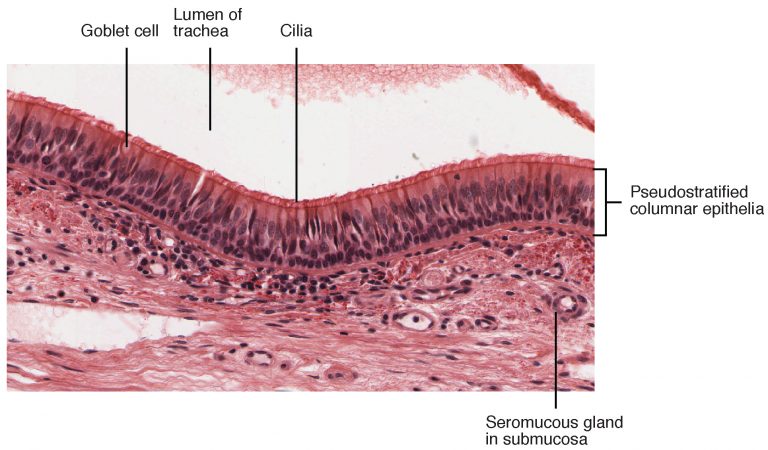
Parts of the pharynx and larynx are lined with stratified squamous epithelium for protection. Most of the conducting portion of the respiratory tract is lined with pseudostratified columnar ciliated cells, also called the respiratory epithelium. This epithelium also contains goblet cells which produce mucus. The mucus and the cilia form the mucocillary escalator which transports foreign particles out of the respiratory tract.
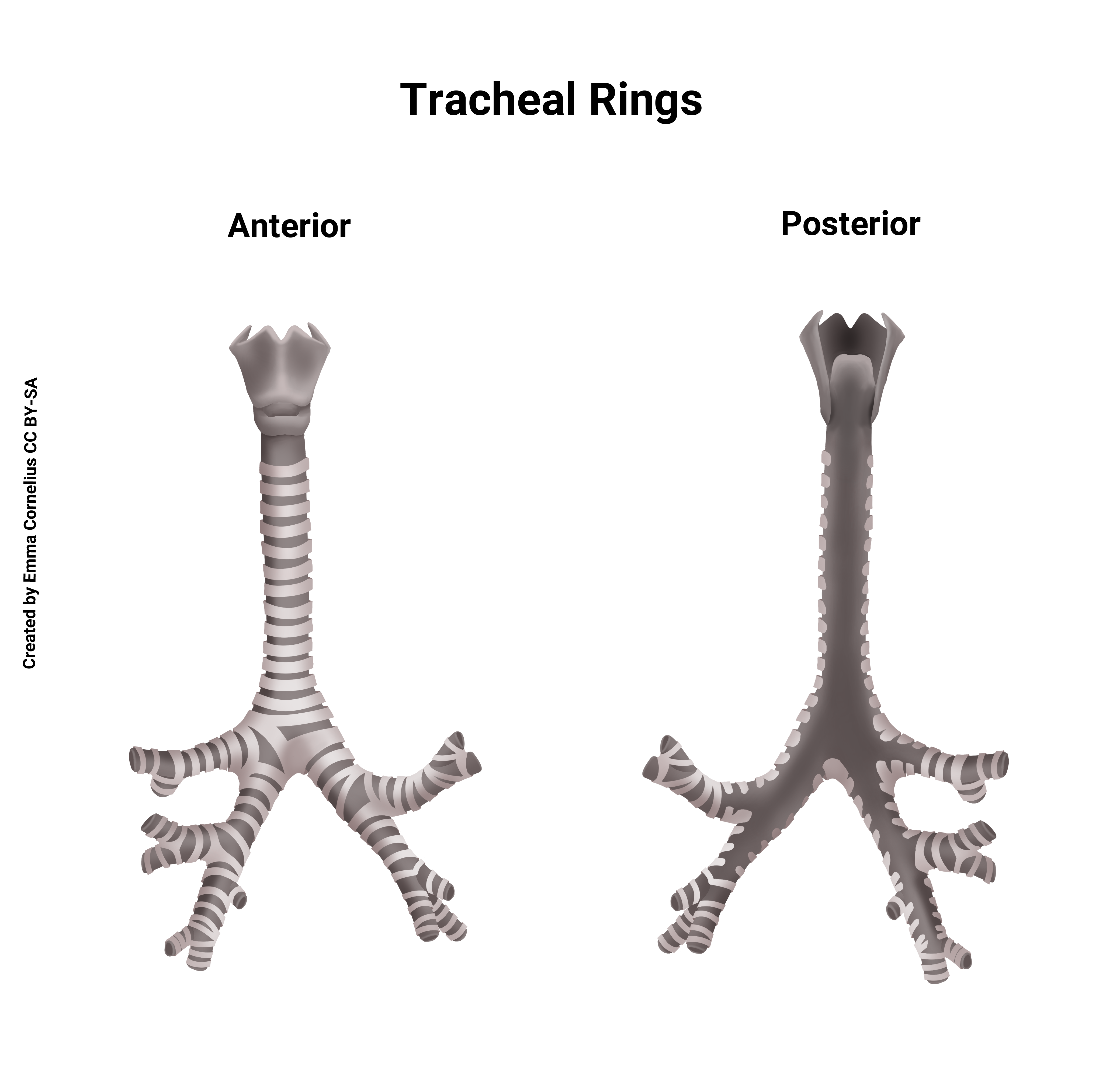
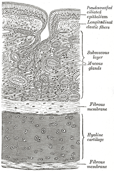
The trachea lies anterior to the esophagus. The trachea is supported by C-shaped rings of hyaline cartilage which prevent the trachea from collapsing and blocking the conduction of air.
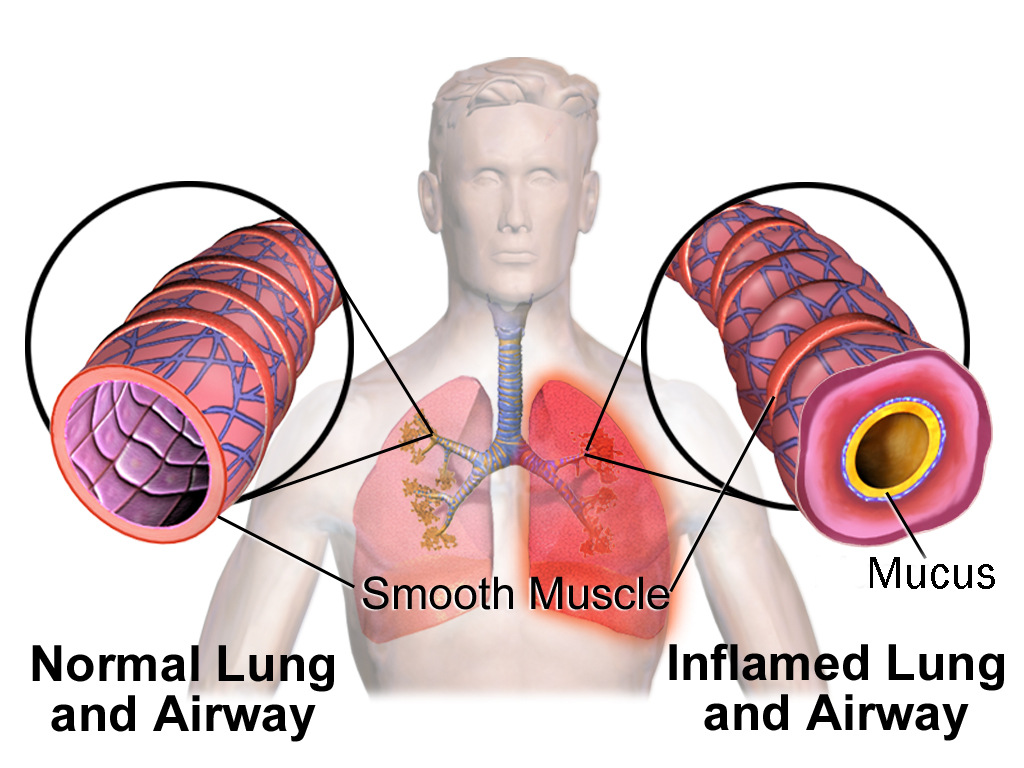
Smooth muscle lines the bronchi and bronchioles. This is important to control the diameter of the airways. When you’re exercising and gasping for air, the sympathetic nervous system stimulates the bronchi and bronchioles to dilate, allowing the passage of more air into the lungs. When you’re sleeping on the couch after your strenuous workout, the airways constrict under parasympathetic stimulation and secretions increase. Inflammatory conditions such as asthma also cause constriction of the airways, trapping air within the lungs.
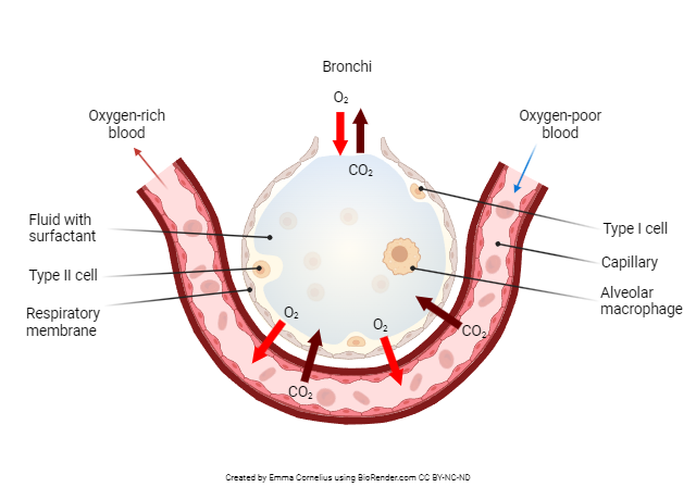
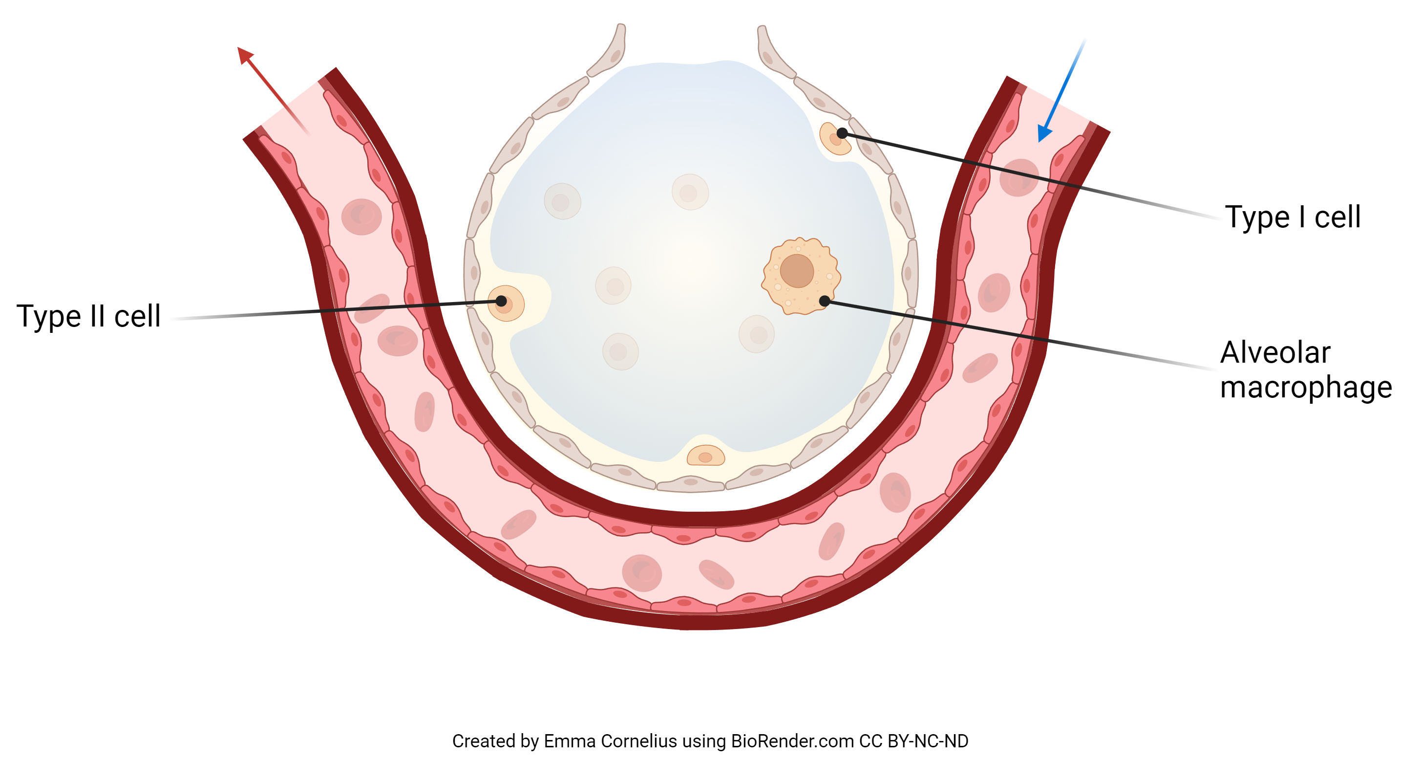
As we move deeper into the respiratory zone, the epithelium changes. Alveoli are lined with two types of epithelium. Type I alveolar cells are simple squamous epithelium. These cells are the site of gas exchange. They are by far the most numerous cell lining the alveoli. The capillaries carrying red blood cells are also lined with a single layer of squamous epithelium. These cells, along with the type I alveolar cells, form the alveolar-capillary (AC) membrane, a thin membrane that allows for easy diffusion of gases. Type II alveolar cells, comprising of simple cuboidal epithelium, secrete surfactant. This is a soap-like substance that decreases surface tension allowing easier inflation of the alveoli and preventing the collapse of alveoli after exhalation. Alveolar macrophages are there for clean up of large particles and invaders.
Media Attributions
- U17-022 Respiratory Epithelium revised © BruceBlaus is licensed under a CC BY (Attribution) license
- U17-021 Pseudostratified Epithelium © Betts, J. Gordon; Young, Kelly A.; Wise, James A.; Johnson, Eddie; Poe, Brandon; Kruse, Dean H. Korol, Oksana; Johnson, Jody E.; Womble, Mark & DeSaix, Peter is licensed under a CC BY (Attribution) license
- U17-023 Tracheal Rings © Cornelius, Emma is licensed under a CC BY-NC-ND (Attribution NonCommercial NoDerivatives) license
- U17-025 Hyaline Cartilage revised © Carter, Henry Vandyke adapted by Burr, Justin is licensed under a CC BY-SA (Attribution ShareAlike) license
- U17-026 Smooth Muscle Airway revised © BruceBlaus is licensed under a CC BY (Attribution) license
- U17-027a Structure of an Alveolus © Cornelius, Emma is licensed under a CC BY-NC-ND (Attribution NonCommercial NoDerivatives) license
- U17-027b Structure of an Alveolus Cell Types Detail © Cornelius, Emma is licensed under a CC BY-NC-ND (Attribution NonCommercial NoDerivatives) license
