Thyroid Gland and Thyroid Hormones
Objective 6
Describe the anatomical location and general structure of the thyroid gland. Identify the general structure of a thyroid follicle and describe the process of thyroid hormone (T3 and T4) synthesis, secretion, transport, and control. Describe the actions of the thyroid hormones, T3 and T4.
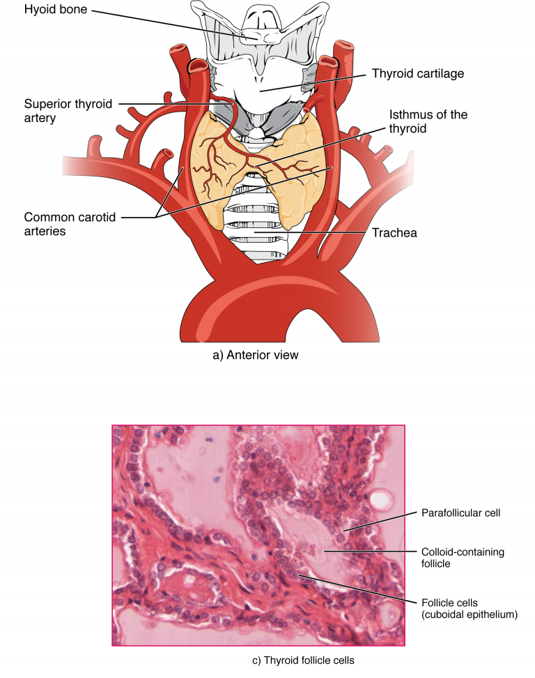
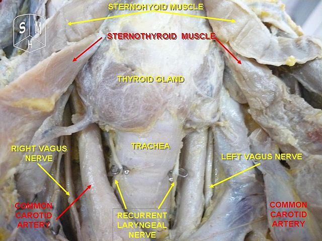 The thyroid gland is easy to find. It is located just inferior to the larynx, specifically the thyroid cartilage (Adam’s apple) and lateral to the trachea. The thyroid gland is butterfly-shaped and consists of two lobes, the left and right. The tissue connection between the two lobes is called the isthmus. Microscopically, the thyroid is made up of small, spherical sacs called thyroid follicles. The follicles make up the largest portion of the gland’s mass.

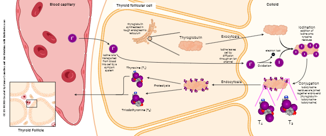
The main functional unit of the thyroid gland is the thyroid follicle. Each follicle consists of a central, open space called the lumen, surrounded by a wall of cells called follicular cells. These cells change shape slightly from more flattened to more tall when stimulated to produce thyroid hormone. A connective tissue basement surrounds the layer of follicular cells.
There is a scattered group of cells surrounding each follicle, parafollicular cells or C cells. The hormone secreted by these cells will be discussed in a later objective.
A thyroid follicle secretes two hormones, thyroxin or tetraiodothyronine (T4) and triiodothyronine (T3). The prefix of each term describes the number of iodine molecules they contain, tetra- meaning four (T4) and tri- meaning three (T3).
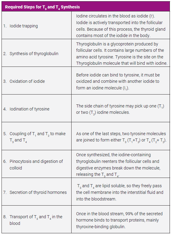
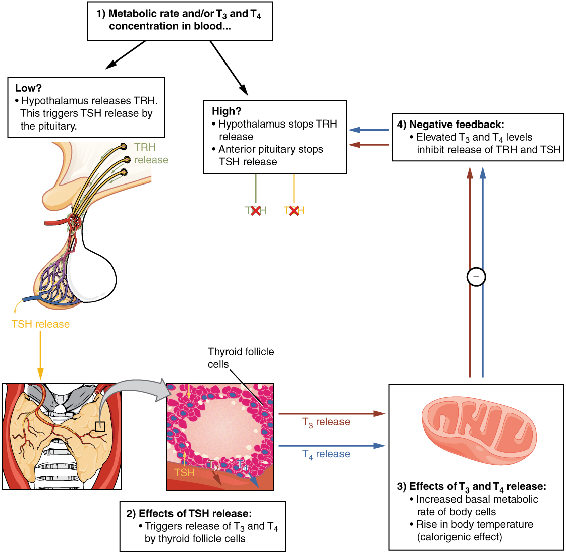
Production of thyroid hormone can be somewhat complex. First, one must understand how to stimulate the thyroid gland. Low T3 and T4 levels, or a low metabolic rate, stimulates the hypothalamus to secrete thyrotropin-releasing hormone (TRH), which results in the anterior pituitary producing thyroid-stimulating hormone (TSH). TSH binds to TSH receptors in the follicular cells and activates a number of processes required to synthesize T3 and T4 (see required steps for T3 and T4 synthesis).
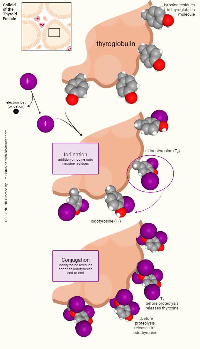
Steps 3 through 5, which occur in the colloid of the thyroid follicle, are shown here in more detail.
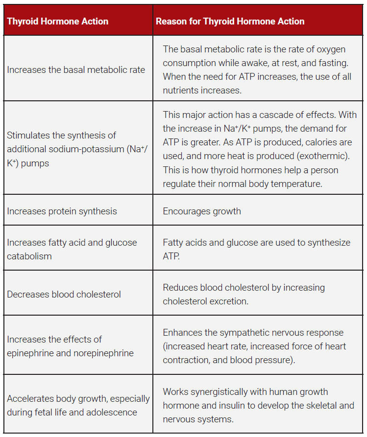
Most of the synthesized thyroid hormone is T4, but T3 is more physiologically potent. However, once secreted, most of the T4 is converted to T3 by enzymatic removal of an iodine. Most body cells have receptors for T3 and T4, so the hormones’ actions are quite broad. It should be noted that the thyroid gland is the only gland to store a large supply of its products (approximately 100 days worth).
Thyroid hormones are regulated in a classic negative feedback loop as illustrated below.
Clinical Connection
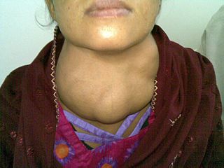 A goiter is simply an enlargement of the thyroid, and it can be found in patients with hypothyroidism, hyperthyroidism, and euthyroidism, which means normal thyroid function. It is important for the physician to determine why the thyroid is enlarged.
In many countries, a goiter is due to iodine deficiency. The thyroid is making every effort to make thyroid hormone, but there isn’t any iodine. In the US, goiters are uncommon because most of the available salt is “iodized”. With the amount of salt consumed in the American diet, iodine deficiency is rare.
Media Attributions
- U14-028 Thyroid Gland © Anatomist90 is licensed under a CC BY-SA (Attribution ShareAlike) license
- U14-029 Thyroid Gland © Biga, Lindsey M.; Bronson, Staci; Dawson, Sierra; Harwell, Amy; Hopkins, Robin; Kaufmann, Joel; LeMaster, Mike; Matern, Philip; Morrison-Graham, Katie; Oja, Kristen; Quick, Devon & Ruyeon, Jon is licensed under a CC BY-SA (Attribution ShareAlike) license
- U14-030 Required Steps for T3 and T4 Synthesis © Newton, Kathy is licensed under a CC BY-SA (Attribution ShareAlike) license
- U14-033c Thyroid Hormone Synthesis © Cornelius, Emma and Hutchins, Jim is licensed under a CC BY-NC-ND (Attribution NonCommercial NoDerivatives) license
- U14-033a Details of Chemical Reactions in the Thyroid Colloid © Hutchins, Jim is licensed under a CC BY-NC-ND (Attribution NonCommercial NoDerivatives) license
- U14-032 Thyroid Hormone Action Table © Newton, Kathy is licensed under a CC BY-SA (Attribution ShareAlike) license
- U14-033b Classic Negative Feedback Loop © Betts, J. Gordon; Young, Kelly A.; Wise, James A.; Johnson, Eddie; Poe, Brandon; Kruse, Dean H. Korol, Oksana; Johnson, Jody E.; Womble, Mark & DeSaix, Peter is licensed under a CC BY (Attribution) license
- U14-034 Goiter © Betts, J. Gordon; Young, Kelly A.; Wise, James A.; Johnson, Eddie; Poe, Brandon; Kruse, Dean H. Korol, Oksana; Johnson, Jody E.; Womble, Mark & DeSaix, Peter is licensed under a CC BY (Attribution) license
