Neurotransmitters
Objective 9
Identify some of the most common neurotransmitters.
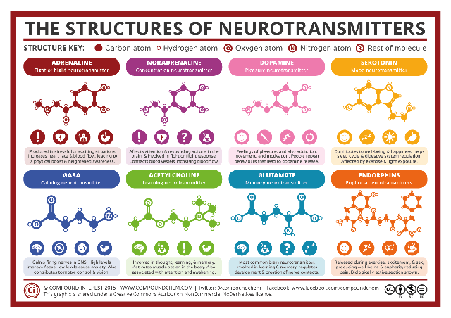
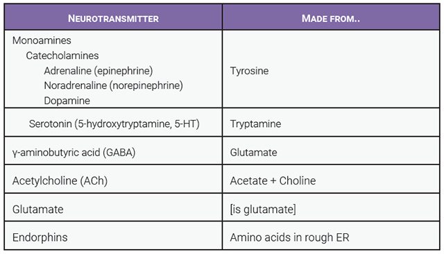
This Compound Interest infographic, and the table, shows eight key neurotransmitters along with their structures. Let’s take some time to learn a little bit about each one.
Adrenaline, Noradrenaline, and Dopamine (Catecholamines)
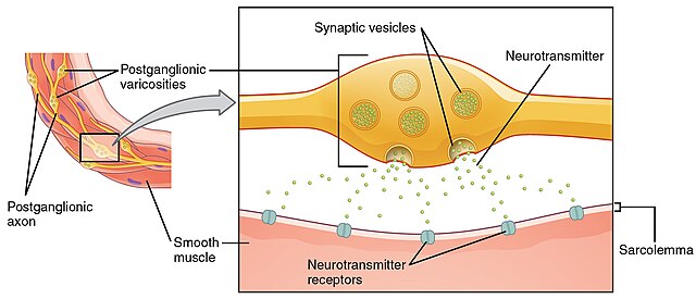
In the autonomic nervous system, catecholamines are released from varicosities, wide spots on the nerve axon. In the central nervous system, they are released from synapses like the ones we studied in Objective 8. The dopaminergic synapse shown below is such an example.
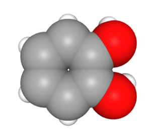
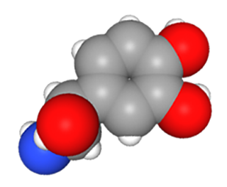
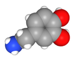
Adrenaline (also called epinephrine), noradrenaline (norepinephrine), and dopamine are collectively called catecholamines. It probably won’t surprise you to know the rings shown, with two hydroxyl groups attached, are called “catechol groups” by chemists, and you’ve already met the amine group in Unit 2. All of these are made from the amino acid tyrosine in a stepwise fashion: first tyrosine makes an intermediate (L-DOPA), then dopamine, then norepinephrine, then epinephrine.
The words “adrenaline” and “epinephrine” both mean the same thing: “chemical from a gland on top of the kidney”, because they were discovered as secretions of the adrenal glands before they were found in the brain. It’s annoying that we have two names for the same chemical and there are all kinds of scientific urban legends about why that is. One thing is for sure, it’s not the fault of your anatomy & physiology professor so don’t blame us.
Adrenaline (epinephrine) is familiar to people with severe allergic responses called anaphylactic shock. They use an “epi pen”, filled with epinephrine, to reverse the effects of another neurotransmitter, histamine. Adrenaline is also familiar to us as a hormone that is released in stressful situations: “when that tiger jumped out at me, I got a shot of adrenaline and ran like the wind”. Anxiety, exercise, love, or surprise can release adrenaline to activate the sympathetic nervous system throughout the body, as we saw in Unit 12 Objective 2.
Noradrenaline (norepinephrine) is the postganglionic neurotransmitter of the sympathetic nervous system. The receptors which bind this neurotransmitter as well as adrenaline are called adrenergic receptors. They come in α and β types. Drugs called β blockers decrease blood pressure by blocking (antagonizing) the increase in blood pressure caused by the adrenaline and noradrenaline binding to the β adrenergic receptor (Unit 16).
Dopamine is important in regulating movement and is diminished in Parkinson disease. It is also a critical part of reward circuitry in the brain.
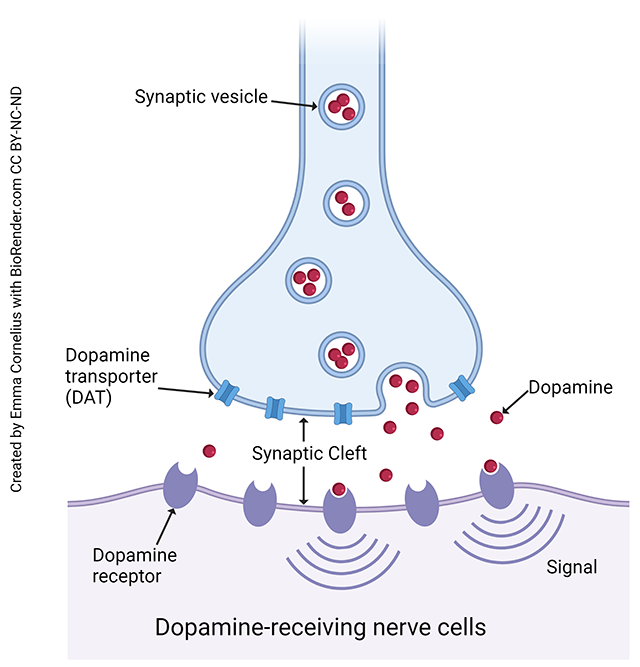
This diagram shows how dopamine is released from synaptic vesicles to cause the generation of a signal in the postsynaptic neuron. We’ll look at the nature of this signal, which is initiated by a G protein-coupled receptor, in Objective 12.
Serotonin
Serotonin (also called by its chemical name 5-hydroxytryptamine or 5-HT) plays an important role in blocking pain transmission in the spinal cord and in regulating mood in the cortex.
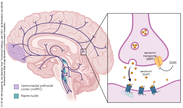
Serotonin is made in the raphe (Latin: “seam”) nuclei of the brainstem, which are so named because they lie close to the midline of the brainstem tegmentum (core) of the midbrain, pons, and medulla. Descending serotonergic systems help limit pain and modify pain perception depending on mood. Ascending serotonergic systems go to various areas of cortex, but psychiatrists have focused on the projections to the ventromedial prefrontal cortex (vmPFC), roughly the same area as the orbitofrontal cortex which we mapped earlier.
Almost all kinds of serotonin receptors are G protein-coupled receptors.
Selective serotonin reuptake inhibitors, such as Prozac©, Celexa©, or Zoloft©, block the reuptake of serotonin and thereby increase the amount of serotonin available at the serotonergic synapse.
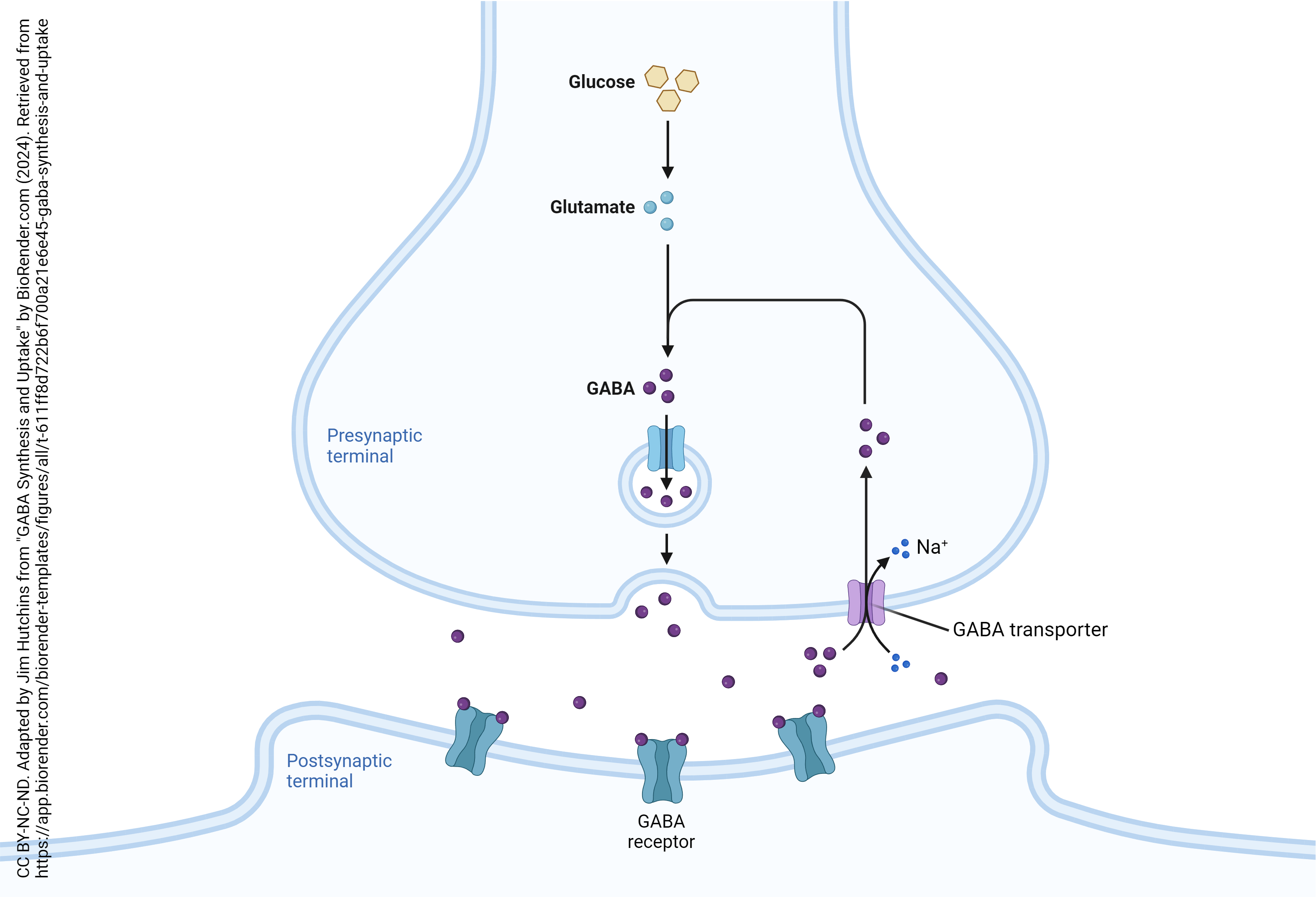
GABA
γ-aminobutyric acid, or GABA, is the most common inhibitory neurotransmitter in the brain. GABA is a slightly modified amino acid which is used at a lot of different synapses. It’s our go-to neurotransmitter when we need inhibition at a synapse. GABA has two types of receptors: GABAA and GABAB. GABAA receptors are a ligand-gated channel also called an ionotropic receptor. When GABA (or a number of different drugs) binds the GABAA receptor, it allows Cl– ions to flow into the neuron, holding it at a voltage below the firing threshold and therefore decreasing the cell’s ability to generate action potentials. GABAA receptors, importantly, are bound by the drugs called benzodiazepines. Xanax® and Valium® are examples of these drugs. The GABAA receptor is also an ethanol receptor. That’s why these two classes of drugs (benzodiazepines and ethanol) don’t mix: in isolation they can produce a fatal CNS depression but in combination the effects are much stronger (synergistic). GABAB receptors are G protein-coupled receptors.
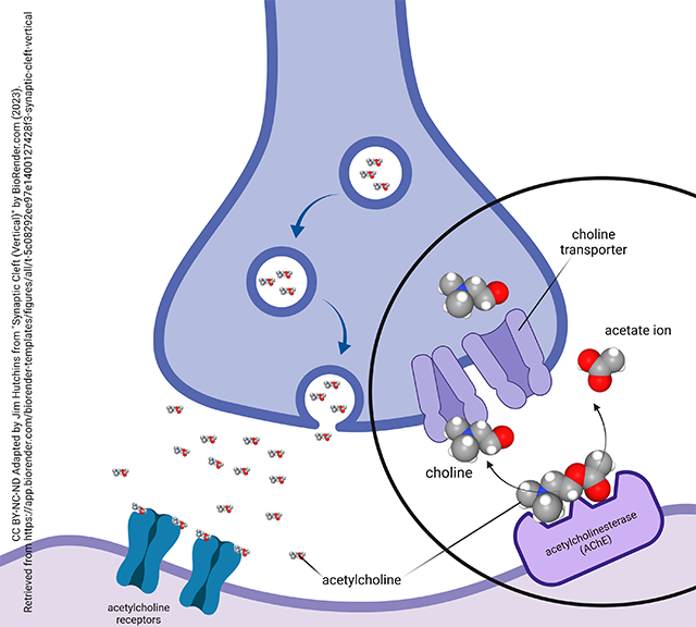
Acetylcholine
 Acetylcholine is just awesome. Dr. Hutchins’ PhD dissertation was called “Evidence for Acetylcholine as a Neurotransmitter in the Human Retina” and you’re welcome to read it. (Just kidding.) Acetylcholine was the first neurotransmitter discovered; the German scientist Otto Loewi called it “vagusstoff” because it was released from the vagus nerve (CN X). We learned about that when we studied the parasympathetic nervous system, but acetylcholine plays a role as a preganglionic neurotransmitter in the sympathetic nervous system as well. Acetylcholine, acting at nicotinic acetylcholine receptors, is the neurotransmitter at the neuromuscular junction. It has fewer roles in the CNS but if you want to know what they are, feel free to request a copy of that dissertation from the Baylor College of Medicine library. You’ll probably be the first.
Glutamate
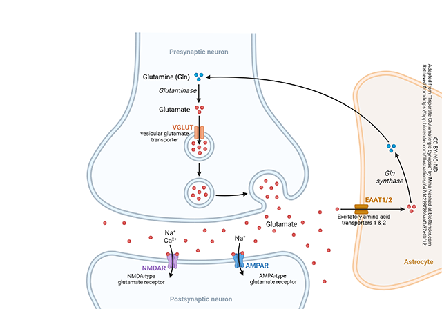 Glutamate is not only a common amino acid, but it also the most abundant neurotransmitter. Working its complicated magic through the n-methyl-d-aspartate (NMDA) receptor, glutamate is the key driver of learning and memory processes as well as many events in the development of the brain. Other types of glutamate receptor have similarly important roles.
Endorphins and Other Neuropeptides
Endorphins are a kind of peptide neurotransmitter: rather than being single amino acids, like glutamate, or modified amino acids, like GABA, these are small strings of amino acids (polypeptides). For example, β-endorphin is 31 amino acids long. β-endorphin binds to the same receptors as morphine, heroin, oxycontin, and other opiates. It is normally released in the brainstem and spinal cord to reduce the pain response.
Over 100 different neuropeptide transmitters are known. These are short amino acid polymers, as short as 3 amino acids (glutamate-histidine-proline, thyrotropin releasing hormone or TRH) and as large as about 100 amino acids.
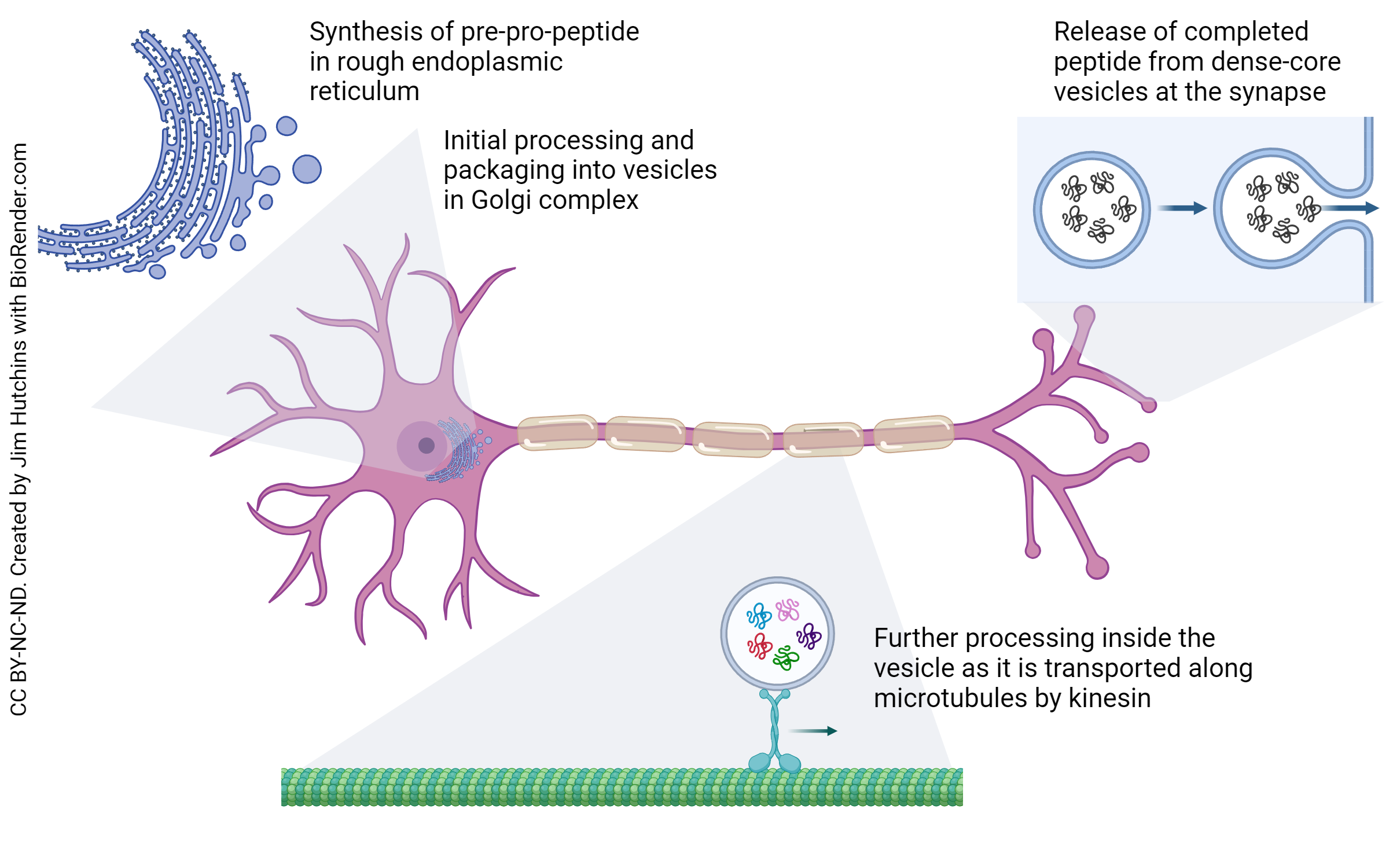
What’s both usual and unusual about neuropeptides is the way they’re made. Like other amino acid polymers, they are made by ribosomes in the rough endoplasmic reticulum (RER) of the neuronal cell body. They are then packaged into vesicles in the Golgi and begin a journey down the axon through a process called anterograde axoplasmic transport. The details have to be seen to be believed, but luckily, in the Canvas course we have a video that shows this process. Kinesin is the motor protein; it is shaped like a little human. It puts the vesicle on its back and then walks down the microtubules which make up the structural core of the axon.
Even stranger perhaps is that the RER makes a huge precursor molecule rather than the peptide itself. This is called a pro-peptide. The Golgi apparatus packs enzymes in the vesicle for the several-day trip to the axon terminal, and over that time, as the vesicle is being carried on kinesin’s back, the enzymes inside are converting the pre-pro-peptide into its final form. For example, the pro-peptide called pro-opiomelanocortin (POMC) makes several different kinds of neuropeptides (also used as hormones). It is named after the most prominent ones: β-endorphin (an endogenous opioid); melanocyte stimulating hormone; and adrenocorticotropic hormone.
Waste products are returned to the cell body for recycling via retrograde axoplasmic transport. This route is also used to send signals from the axon terminal to the cell body.
Neurosteroids
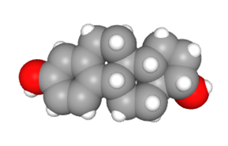
Lipid signaling molecules based on the structure of cholesterol are called steroids. Several dozen of these steroid signaling molecules are active in the nervous system and are therefore called neurosteroids. We’ve already seen, and you probably know, a couple of these: the female sex hormone 17-β-estradiol (one of the estrogens) and the male sex hormone testosterone. The stress hormone cortisol is another example.
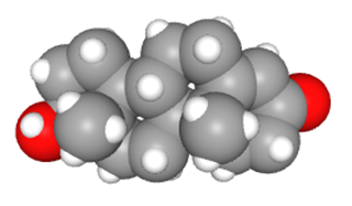
Many people find that these hormones alter their brain functions sometimes in good ways, sometimes not. When these neurosteroids are released into the bloodstream to have an effect on non-neural tissue, we call them hormones and discuss them in Unit 14.
Media Attributions
- U13-067 Chemical Structures of Neurotransmitters © Compound Interest is licensed under a CC BY-NC-ND (Attribution NonCommercial NoDerivatives) license
- U13-068 Table of Neurotransmitters © Hutchins, Jim and Bizzell, Lizz is licensed under a CC BY-SA (Attribution ShareAlike) license
- U13-068a Autonomic Varicosities © Betts, J. Gordon; Young, Kelly A.; Wise, James A.; Johnson, Eddie; Poe, Brandon; Kruse, Dean H. Korol, Oksana; Johnson, Jody E.; Womble, Mark & DeSaix, Peter is licensed under a CC BY (Attribution) license
- U13-069 Catechol Conformer3D © PubChem is licensed under a CC0 (Creative Commons Zero) license
- U13-071 Norepinephrine Conformer © PubChem is licensed under a CC0 (Creative Commons Zero) license
- U13-070 Dopamine Conformer3D © PubChem is licensed under a CC0 (Creative Commons Zero) license
- U13-076 Dopamine © Cornelius, Emma is licensed under a CC BY-NC-ND (Attribution NonCommercial NoDerivatives) license
- U13-078a Serotonin Pathways and SSRIs © Hutchins, Jim is licensed under a CC BY-NC-ND (Attribution NonCommercial NoDerivatives) license
- U13-077 GABA Synapse © Hutchins, Jim is licensed under a CC BY-NC-ND (Attribution NonCommercial NoDerivatives) license
- U13-072 Dissertation © Hutchins, Jim is licensed under a CC BY-SA (Attribution ShareAlike) license
- U13-065 Acetylcholinesterase at the Cholinergic Synapse © Hutchins, Jim is licensed under a CC BY-NC-ND (Attribution NonCommercial NoDerivatives) license
- U13-075 Glutamatergic Synapse © Hutchins, Jim and Nashed, Mina is licensed under a CC BY-NC-ND (Attribution NonCommercial NoDerivatives) license
- U13-078 Axonal Transport of Neuropeptides © Hutchins, Jim is licensed under a CC BY-NC-ND (Attribution NonCommercial NoDerivatives) license
- U13-079 Estradiol Conformer © PubChem is licensed under a CC0 (Creative Commons Zero) license
- U13-080 Testosterone Conformer © PubChem is licensed under a CC0 (Creative Commons Zero) license
