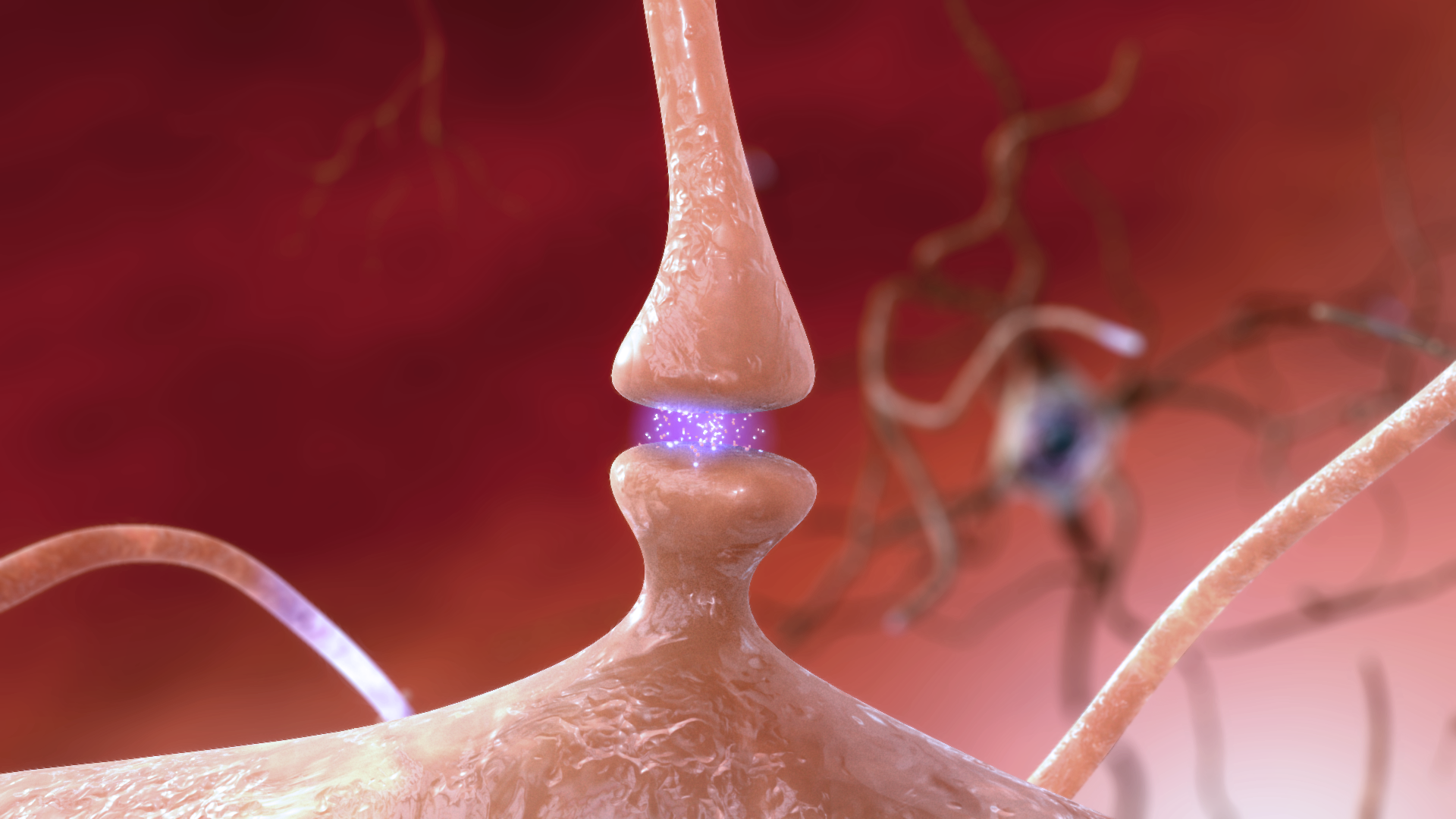
The Synapse
Objective 8
Explain how a synapse works.
 In Objectives 4-7, we saw the involvement of voltage-gated sodium and potassium channels in the creation and propagation of the action potential. Now, we turn our focus to a third kind of voltage-gated channel, the voltage-gated calcium (Ca2+) channel. This channel predominates where there are signals being sent from one neuron to a target cell, so we think of them as being associated with the axon terminals. Because there are 10,000 times as many calcium ions outside the cell as inside, the concentration gradient overwhelms any electrical gradient that might exist. If a voltage-gated Ca2+ channel opens, Ca2+ will always rush into the neuron.
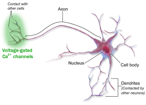 Voltage-gated Ca2+ channels are found only in the active zone. Active zones are, in turn, found only on the sending side of a synapse, a contact between a nerve cell and a target cell. The side sending the information is called the presynaptic side, and the side receiving the information is called the postsynaptic side. The postsynaptic target cell can be another neuron; skeletal, cardiac, or smooth muscle; or a gland. We’ve already seen skeletal muscle as a target when we studied the neuromuscular junction in Unit 10. When the target is distant from the presynaptic neuron and the chemical signal is carried by the bloodstream, it’s called a hormone, and we’ll study those in Unit 14.
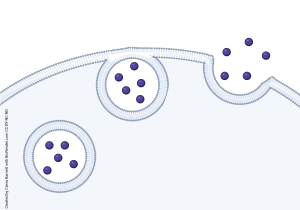 The release of a neuron’s chemical signal (neurotransmitter) from the presynaptic terminal is just a special case of a process we described in Unit 4, exocytosis (exo–, “out of”; –cyt–, “cell”; –osis, “process”). Here, we’re moving neurotransmitter out of the cell in response to an intracellular signal. The neurotransmitter is contained within vesicles (Latin: “little bladder”).
As you might imagine, to release neurotransmitter on demand, and to receive the signal carried by the neurotransmitter, requires a complicated and exquisite machinery.
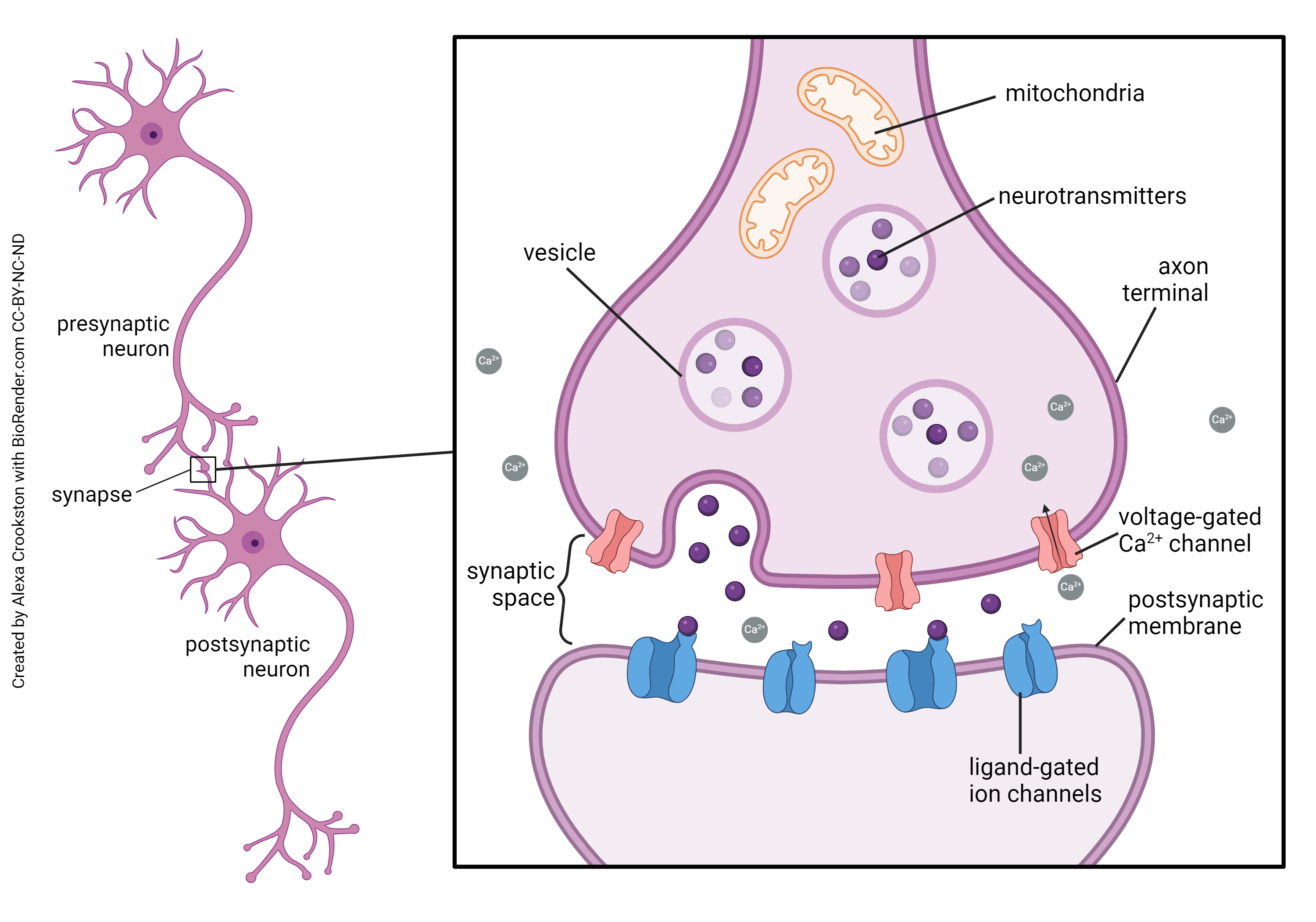
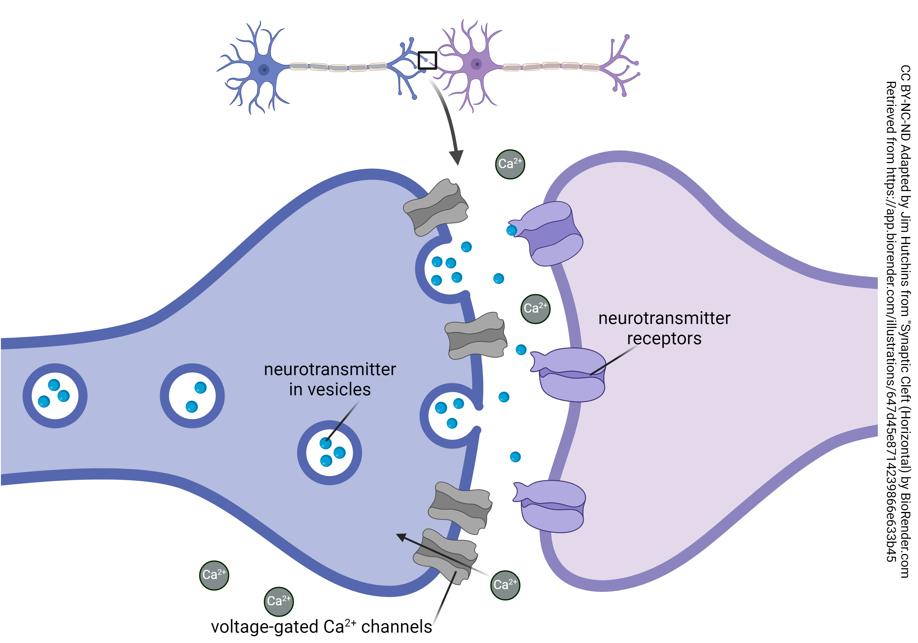
This diagram shows the parts of a synapse. The presynaptic neuron and postsynaptic neuron are separated by a synaptic cleft. A voltage change causes the opening of voltage-gated Ca2+ channels. This, in turn, leads to the release of neurotransmitter from vesicles. The neurotransmitter diffuses across the synaptic cleft and interacts with ligand-gated ion channels on the postsynaptic membrane.
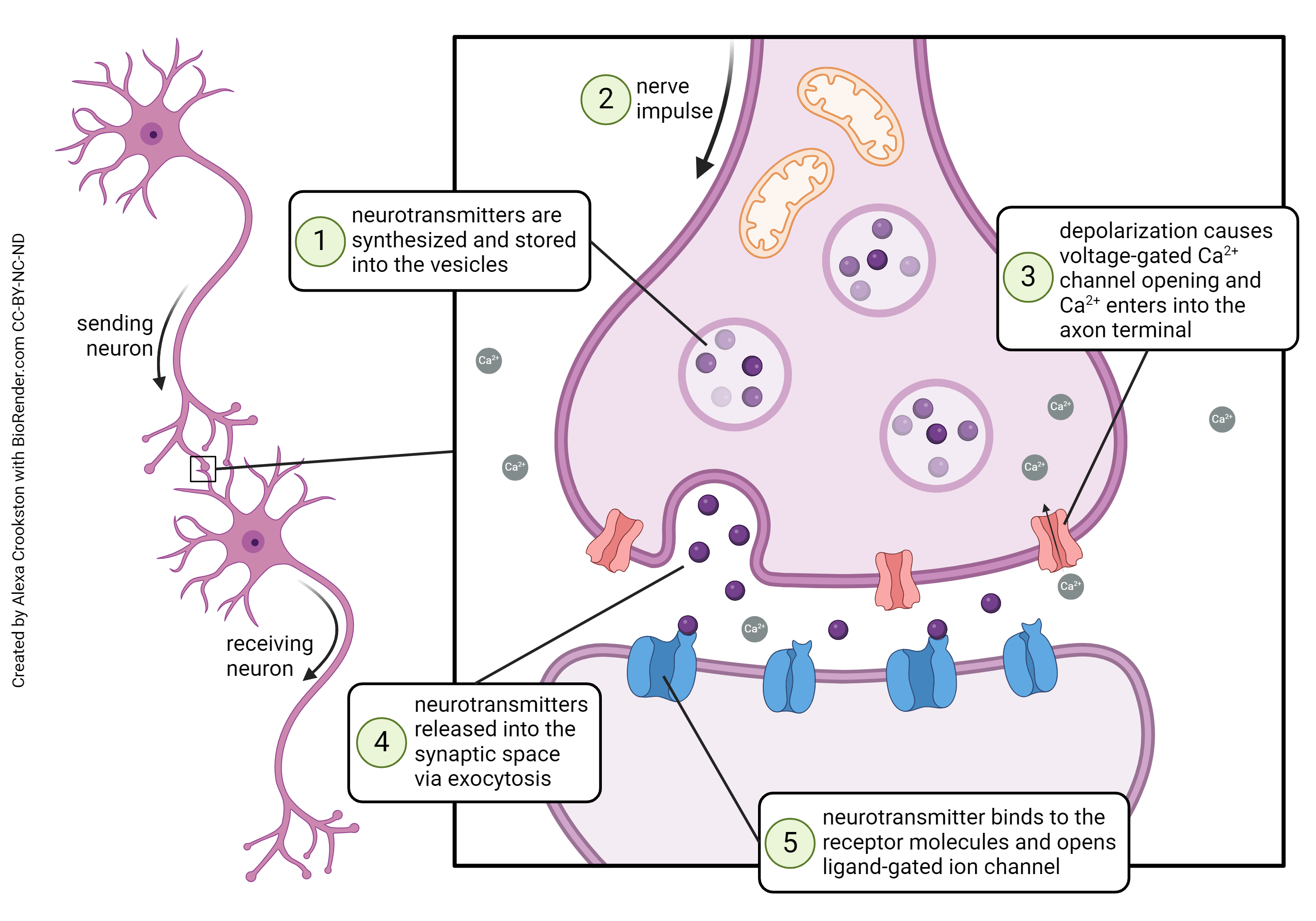
Here are the steps, in order:
- the action potential, or even a smaller voltage change called a graded potential, arrives at the axon terminal and opens voltage-gated Ca2+ channels;
- Ca2+ enters the presynaptic terminal and, through a complicated process involving dozens of steps, triggers exocytosis;
- the vesicles containing neurotransmitter spill their contents into the synaptic cleft, and the neurotransmitter diffuses across the synaptic cleft;
- where it encounters transmembrane protein receptors, which transduce the chemical signal into an
- electrical signal changing the voltage of the postsynaptic cell or
- a biochemical change changing the function of the postsynaptic cell or electrical signal in the postsynaptic cell.
On the postsynaptic side, a group of cytoskeletal and related proteins form the postsynaptic density, which appears as a “dark fuzz” under the electron microscope.
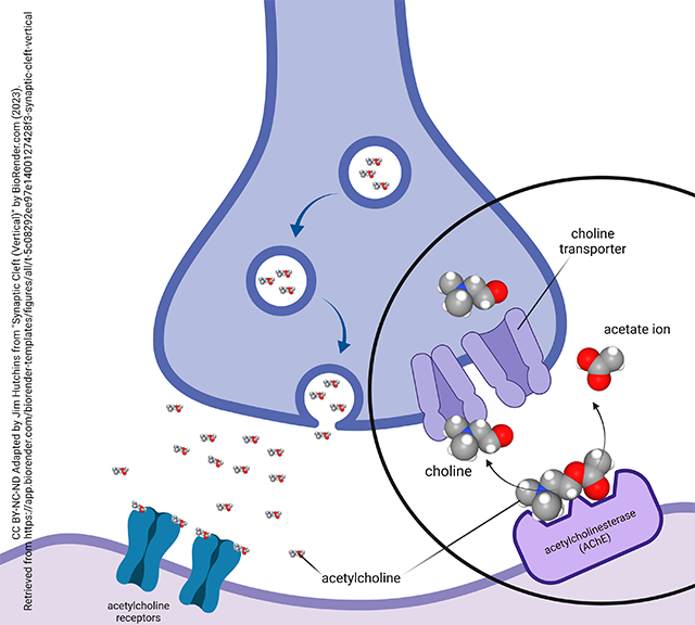
One aspect of neurotransmission we haven’t yet considered is the focus of this diagram: removal of neurotransmitter from the synapse.
Neurotransmitter removal is important because each neurotransmitter molecule can activate a receptor, release from the receptor, and then activate the same receptor or another. This happens until the neurotransmitter is removed.
There are three mechanisms for removing neurotransmitter. Some neurotransmitters use one of these, some two, and some a combination of all three.
Degradation enzyme. For example, acetylcholine is broken down by the enzyme acetylcholinesterase (AChE).
Transporter. These are symport or antiport systems that move the neurotransmitter back into the presynaptic terminal, removing its access to receptors. A drug that blocks these transporters therefore makes more of the neurotransmitter available and increases the influence of the transmitter. An example would be Prozac®, Lexapro®, Paxil®, Zoloft®, and Celexa®, all brand names for selective serotonin reuptake inhibitors (SSRIs). By blocking the serotonin transporter, this class of drugs increases the serotonin binding to receptors.
Metabolizing enzyme. Where a degradation enzyme works extracellularly to inactivate the neurotransmitter, a metabolizing enzyme works intracellularly to break down the neurotransmitter so its pieces can be used to synthesize new transmitter later. An example of this would be monoamine oxidase (MAO), which is found on mitochondria and breaks down monoamine neurotransmitters.
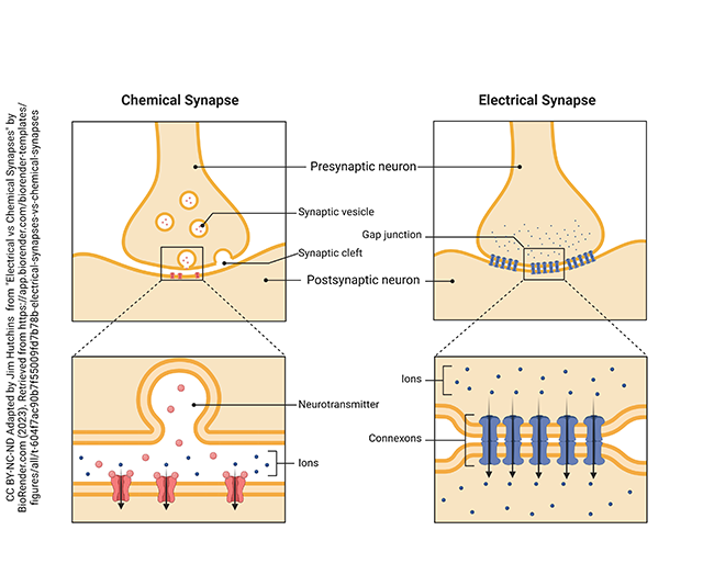 Some synapses do not utilize the complicated machinery involved in the vesicular release of transmitter and the presence of postsynaptic receptors. Rather, these electrical synapses use gap junctions (as introduced in Unit 7) to pass ions directly between neurons. Like gap junctions, electrical synapses consist of cells joined by structures called connexons which are made up of a protein called connexin. When the cell at the top depolarizes, then the ions that flow into the presynaptic cell can continue diffusing into the postsynaptic cell through the connexon proteins of the electrical synapse.
Media Attributions
- U11-025 Synapse © National Institute on Aging, National Institutes of Health is licensed under a CC BY-NC (Attribution NonCommercial) license
- U13-060 Voltage Gated Ca Channel Distribution © BruceBlaus adapted by Jim Hutchins is licensed under a CC BY (Attribution) license
- U13-062 voltage gated calcium channels at the synapse © Jim Hutchins | BioRender is licensed under a CC BY-NC-ND (Attribution NonCommercial NoDerivatives) license
- U13-061 Cell Membrane in Exocytosis © Barnett, Cierra Memphis is licensed under a CC BY-NC-ND (Attribution NonCommercial NoDerivatives) license
- U13-063 Neurotransmitter System (anatomy) © Crookston, Alexa is licensed under a CC BY-NC-ND (Attribution NonCommercial NoDerivatives) license
- U13-064 Neurotransmitter System (action) © Crookston, Alexa is licensed under a CC BY-NC-ND (Attribution NonCommercial NoDerivatives) license
- U13-065 Acetylcholinesterase at the Cholinergic Synapse © Hutchins, Jim is licensed under a CC BY-NC-ND (Attribution NonCommercial NoDerivatives) license
- U13-066 Comparison of Electrical vs Chemical Synapses © Hutchins, Jim is licensed under a CC BY-NC-ND (Attribution NonCommercial NoDerivatives) license
