Sensory Systems | Visual (Sight) System
Objective 7
Label the components of the eye. List the cell types of the retina. Trace the pathway taken by neural information as it passes from retina to brain.
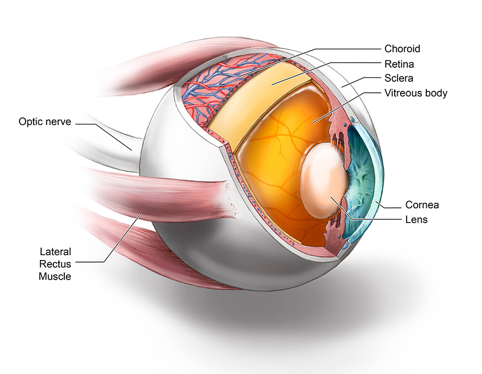 The eye has a number of structures specialized for focusing an image on the retina, which is the receptive sheet of the eye (more about that later).
The eye is enclosed in an orbit, a bony structure that is also filled with fat (adipose) cells to cushion and support the eye.
It is moved by six extraocular muscles.
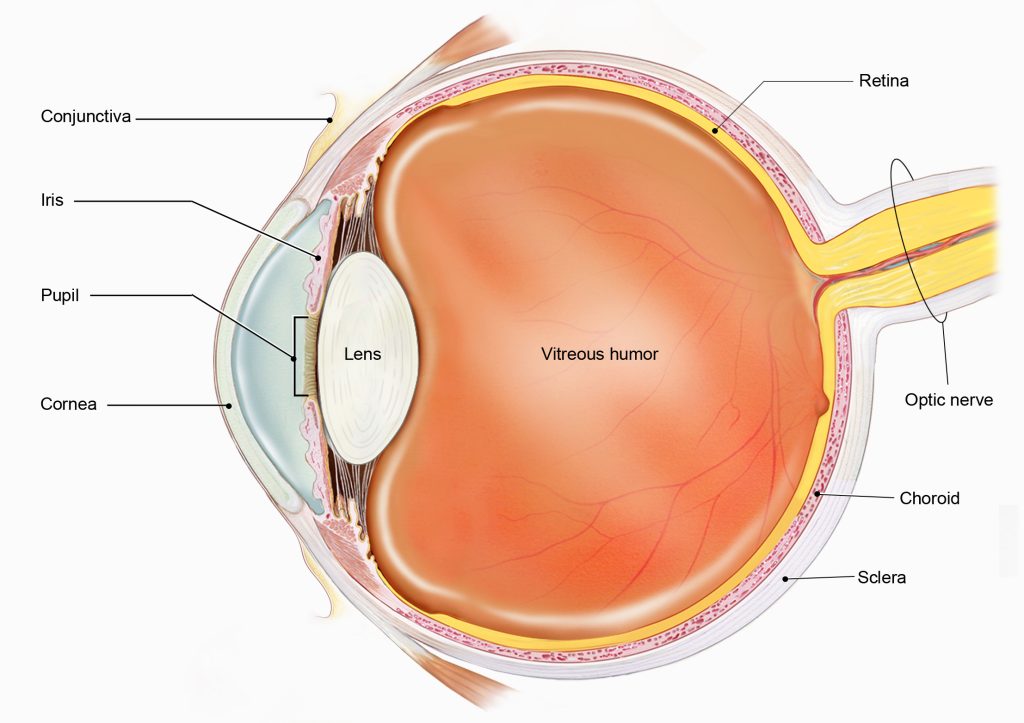
A thin mucous membrane, the conjuctiva, covers the sclera and is also continuous with the internal surfaces of the upper and lower palpebrae. When the conjuctiva becomes inflamed, the patient has conjuctivitis, often called “pink eye”. This can result from either a bacterial or viral infection.
The eye is divided into three tunics, which are related embryologically and also have clinical importance.
From outermost to innermost, the three tunics and their constituents are:
- The fibrous tunic, consisting of the sclera (“white of the eye”) and cornea (clear covering over the pupil). In both eyeball images shown here, the fibrous tunic is white or blue.
- The vascular tunic, also called the uvea, consisting of the iris (colored part of the eye surrounding the pupil); the ciliary body (controls the shape of the lens) and the choroid (choriocapillaris; supplies blood to the retina and other structures of the eye). When this entire layer is inflamed it is called uveitis and may threaten the patient’s sight. Structures comprising the uvea are shown in pink in these diagrams.
- The nervous tunic, consisting of the retina, an outpocketing of the CNS inside the eye. The nervous tunic or neural retina is shown in yellow in these drawings of the eye.
Two viscous (i.e. goopy) liquids help maintain the shape of the eyeball. The vitreous body is the jelly-like substance in contact with the retina in the larger, central chamber of the eye. The other liquid is called the aqueous humor.
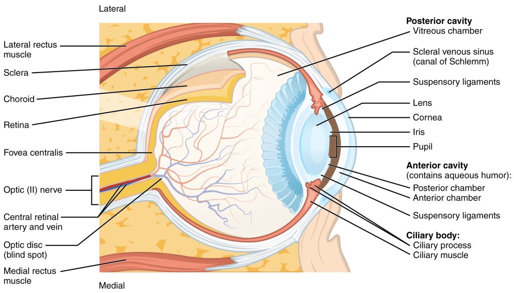
The ciliary body filters blood to secrete aqueous humor into the anterior chamber, located between the lens and inside of the cornea (blue in this diagram). Aqueous humor is a filtrate of the blood that nourishes the lens and cornea. These structures have no blood supply, because blood vessels would destroy the optical properties of these clear structures.
Aqueous humor is made by the ciliary body and flows along the posterior surface of the iris in the narrow posterior chamber. It then flows through the pupil and fills the anterior chamber. The scleral venous sinus (the old name is much more fun to say: the “canal of Schlemm”) is a drainage system for the aqueous humor.
The lens is a clear structure, shaped like an M&M®, that works with image on the retina. When the eye is focused on a distant object, the cornea to focus an image on the retina pulls on suspensory ligaments (zonular fibers; old name the “Zonules of Zinn” — learning anatomy used to be a lot more fun) to flatten the lens (regular M&M®). When the eye is focused on a near object, the ciliary muscle relaxes, and the lens snaps back into a more spherical shape (peanut M&M®). This process, which changes the shape of the lens to focus on objects near and far, is called accommodation.
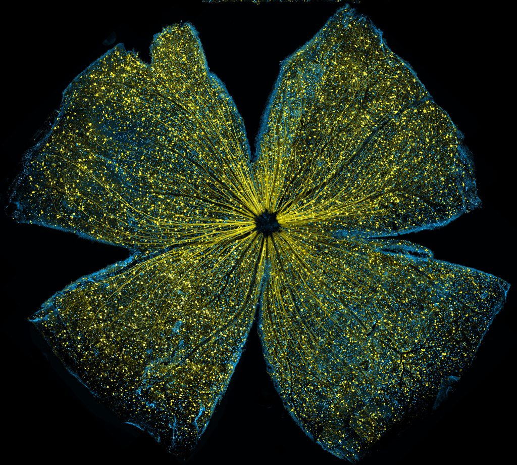
The retina is a thin sheet of photoreceptors, processing cells, and output cells. Oddly, the processing cells and output cells are on top of the photoreceptors, so that light entering the eye has to pass through all the other cell types before it reaches the photoreceptors. Thus the layer of photoreceptor cell bodies is called the outer nuclear layer (ONL), because it’s furthest from the center of the eyeball; the layer of processing cell bodies is called the inner nuclear layer (INL); and the layer of ganglion cell bodies is called, naturally enough, the ganglion cell layer.
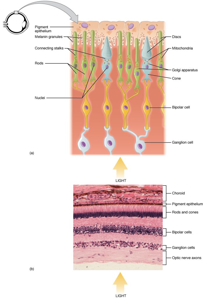
There are five types of cells in the retina, with three functions: transduction; processing; and output.
Transduction (turning photons into a neural signal)
- photoreceptors
-
- rods
- cones
Processing
- horizontal cells
- bipolar cells
- amacrine cells
Output
- ganglion cells
- M-type specialized for motion
- P-type specialized for details
- K-type (also called non-M, non-P) specialized for color vision
There are two kinds of neural processing going on in the retina. There is a straight-through pathway that passes information from photoreceptors to bipolar cells to ganglion cells. The information that passes in this way pretty much runs perpendicular to the retinal sheet.
There is also a lateral pathway that sends information sideways, so that neighboring photoreceptors have a way of comparing information and neighboring bipolar cells can do the same. That’s not shown here, and it’s not essential for you to know.
Central Visual Pathways
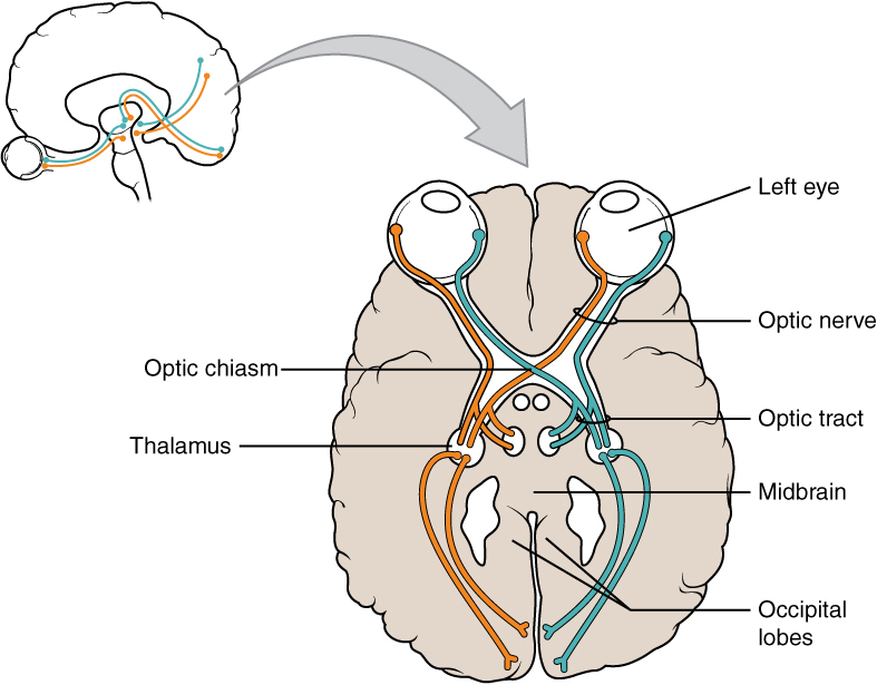
Visual information leaves the retina on ganglion cell axons. These pass out of the eye and form the optic nerve (CN II). Photons from the left side of the world hit the left retina nearest the nose, and hit the right retina nearest the temples.
Therefore, axons from the left nasal retina and right temporal retina, which carry information from photons in the left half of the world, join together at the optic chiasm and form the right optic tract. The right optic tract ends in the right side of the thalamus.
Photons from the right half of the world strike the right nasal retina and the left temporal retina. The ganglion cell axons from the left temporal retina stay on the same side while the axons from the right nasal retina switch sides at the optic chiasm to form the left optic tract. These axons terminate in the left thalamus.
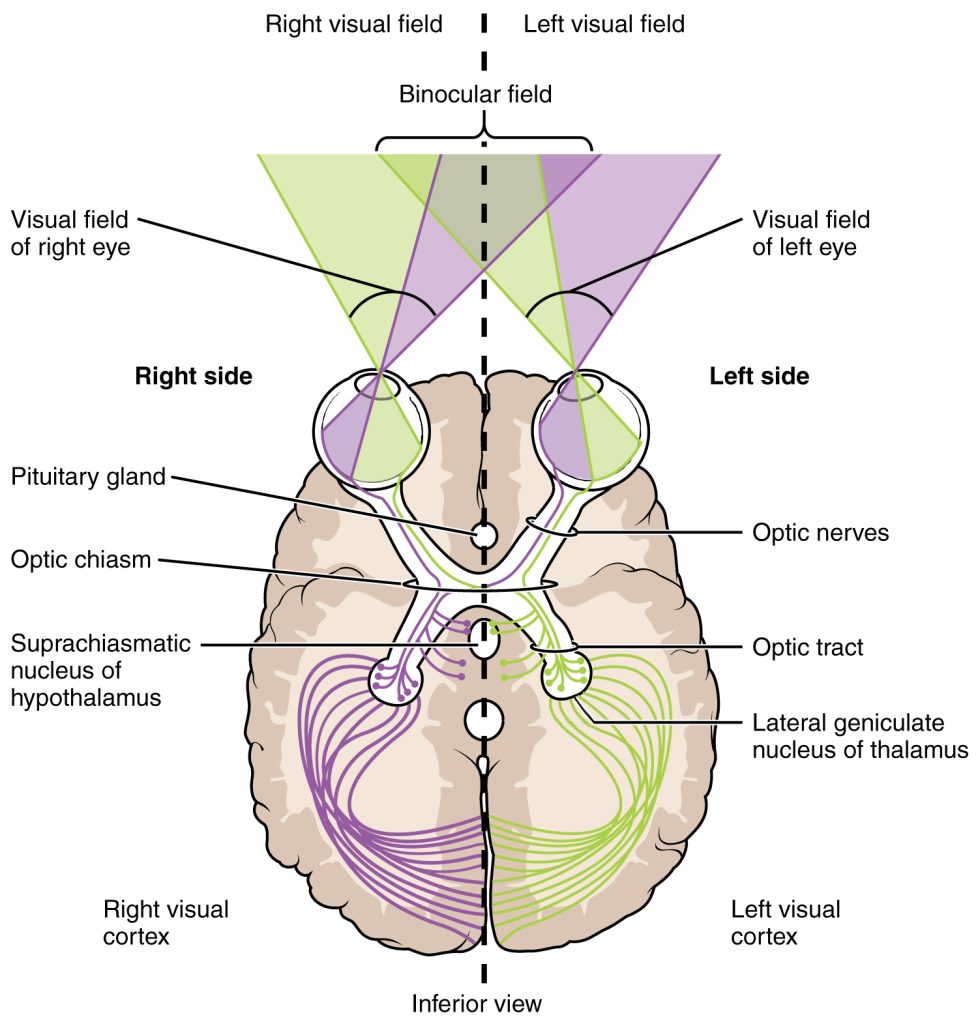
The thalamic relay nucleus is called the lateral geniculate nucleus (LGN). The left LGN contains six complete maps (representations) of the right side of the world; the right LGN contains six complete maps of the left side of the world. In this way, as we expect, sensory information decussates (crosses) at the optic chiasm so that one side of the world now occupies neurons on the opposite side of the brain.
The pathway between the LGN and visual cortex is called the optic radiations. There is a further anatomical separation of the visual maps but we will not discuss that here.
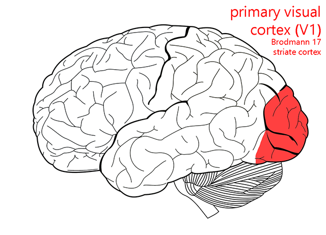
 The primary visual cortex has a lot of names. Unfortunately, we have to learn them all, because different authorities use them interchangeably and without apparent purpose.
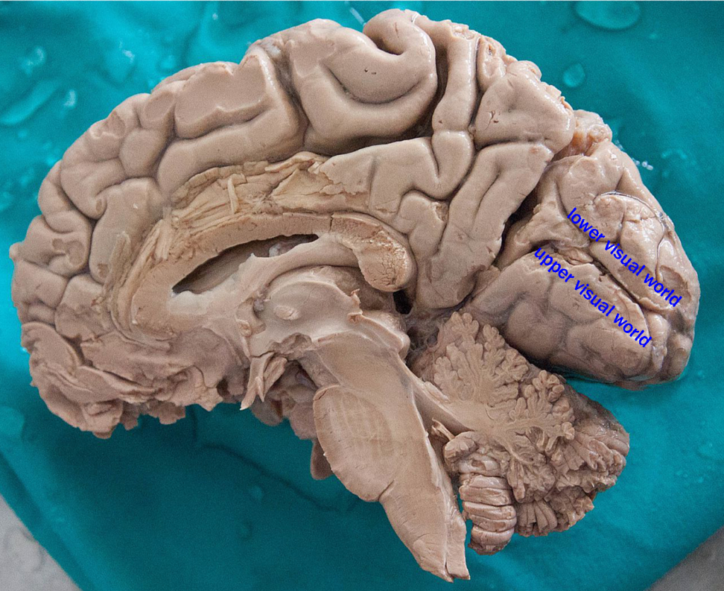
Regardless of what we call it, the visual cortex is on either bank of the calcarine sulcus, a prominent groove in the occipital lobe that we can only see in a midsagittal cut. The macular region, representing the central 5° of vision, takes up about ¼ to ½ of the “real estate” in the visual cortex. This number varies between individuals, and it’s tempting to speculate on how their visual perception might change as a result.
At each level of the visual system, from the retina to the LGN to V1 and beyond, into the visual association areas (Brodmann 18 and 19) of cortex, there are visuotopic or retinotopic maps: two adjacent points in visual space, like your index and middle finger if you hold your hand out and look at it, are represented by two adjacent cells in the retina, LGN, and cortex.
Media Attributions
- U12-064 Eye Cutaway © Kissinger, Ryan \ National Institute of Allergy and Infectious Diseases, NIH is licensed under a Public Domain license
- U12-065 Eye Cross Section © National Eye Institute, National Institutes of Health adapted by Hutchins, Jim is licensed under a CC BY-NC (Attribution NonCommercial) license
- U12-066 Visual Eye Cross Section © Betts, J. Gordon; Young, Kelly A.; Wise, James A.; Johnson, Eddie; Poe, Brandon; Kruse, Dean H. Korol, Oksana; Johnson, Jody E.; Womble, Mark & DeSaix, Peter is licensed under a CC BY (Attribution) license
- U12-067 Mouse Retina © Kim, Kenyoung; Ju, Wonkyu and Ellisman, Mark, National Center for Microscopy and Imaging Research, University of California, San Diego is licensed under a CC BY-NC (Attribution NonCommercial) license
- U12-068 Visual Retina © Betts, J. Gordon; Young, Kelly A.; Wise, James A.; Johnson, Eddie; Poe, Brandon; Kruse, Dean H. Korol, Oksana; Johnson, Jody E.; Womble, Mark & DeSaix, Peter is licensed under a CC BY (Attribution) license
- U12-069 Optic Nerve © Betts, J. Gordon; Young, Kelly A.; Wise, James A.; Johnson, Eddie; Poe, Brandon; Kruse, Dean H. Korol, Oksana; Johnson, Jody E.; Womble, Mark & DeSaix, Peter is licensed under a CC BY (Attribution) license
- U12-070 Visual Fields Corrected © Betts, J. Gordon; Young, Kelly A.; Wise, James A.; Johnson, Eddie; Poe, Brandon; Kruse, Dean H. Korol, Oksana; Johnson, Jody E.; Womble, Mark & DeSaix, Peter is licensed under a CC BY (Attribution) license
- U12-071 Primary Visual Cortex © Carter, Henry Vandyke adapted by Jim Hutchins and Alexa Crookston is licensed under a CC BY-SA (Attribution ShareAlike) license
- U12-072 Midsagittal Human Brain Dissected © Gümüş, Hikmet adapted by Hutchins, Jim is licensed under a CC BY-SA (Attribution ShareAlike) license
