Diffuse Systems of the Brain
Objective 10
Report on the diffuse systems of the brain, including those for emotion, homeostatic control, and arousal.
The Limbic System (Limbic Lobe)
The limbic system, also called the limbic lobe (Latin: limbus, “border, fringe”), plays a key role in emotional behavior. First named “le grand lobe limbique” by Paul Broca, the components and wiring of the limbic system were worked out by James Papez in 1937. The limbic system forms a border or fringe that encircles the brainstem and thalamus.
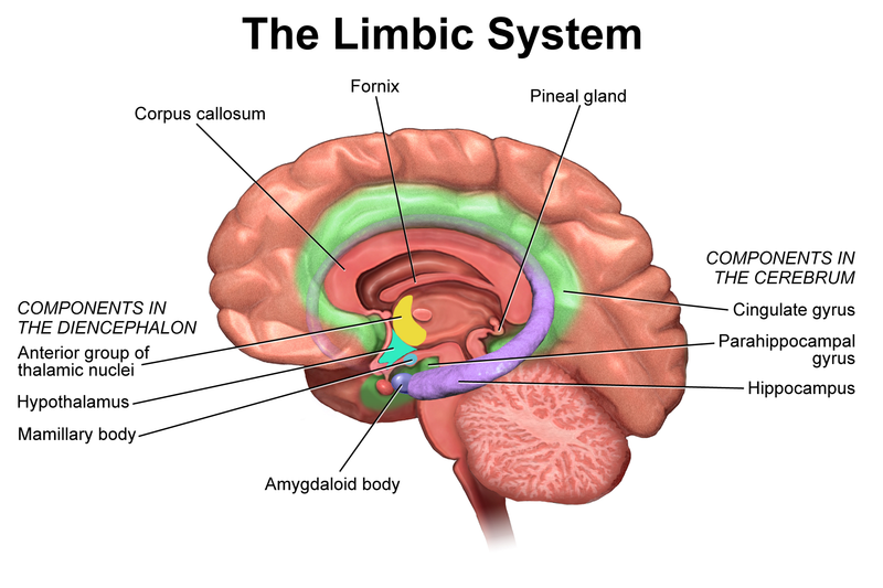
In the original circuit proposed by Papez, the five structures of the limbic lobe (hippocampus, mammillary bodies, anterior nucleus of the thalamus, cingulate gyrus and parahippocampal gyrus) were connected in a circular pathway. The fornix connects the hippocampus to the mammillary bodies. The mammillothalamic tract connects the mammillary bodies to the anterior nucleus of the thalamus. The other pathways we won’t name.
Nice to know but not essential: More recent work has added the amygdala, olfactory bulb, stria medullaris and stria terminalis to the limbic lobe.
All of the structures listed are important in emotional behaviors. The olfactory bulb which processes smell (olfactory) information is an important part of emotion. Try to smell burning flesh, burning food, or feces, without having an emotional response to the odor.
The hippocampus is involved in learning and memory. It makes sense: if you experience a very dangerous situation, you want to remember what led to it, so you can avoid it in the future. This circuit also shows us how important emotion is when we want to learn something. In the absence of emotion, it is very difficult to activate this circuit, and the ability to learn is impaired. We will see the central learning and memory function of the hippocampus in a later objective.
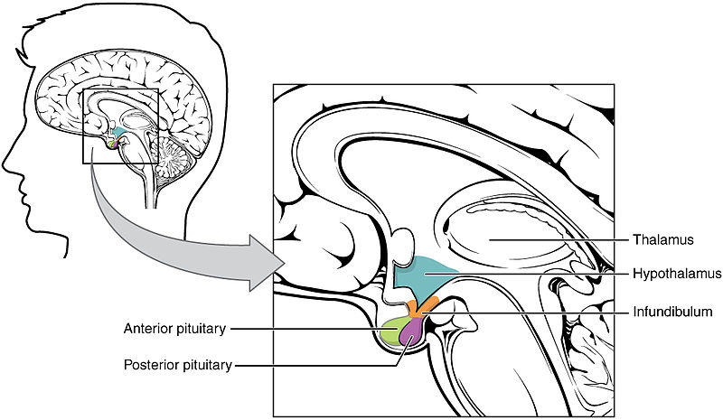 The hypothalamus, as the name suggests, is located just underneath the thalamus. It is intimately associated with the pituitary gland.
 We will look at the hypothalamus and pituitary gland in some detail in Unit 14 when we discuss the endocrine system. For now, we’ll just cover some general anatomy and physiology.
Within the hypothalamus are dozens of small nuclei, clusters of brain cells that are responsible for a particular function.
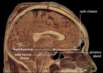
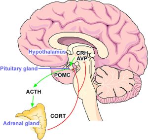 The hypothalamus is the main control center for body homeostasis, as described in Unit 1. For example, body temperature (both “heating” and “cooling”), sexual function, eating, drinking, and emotional state (angry, sad, happy) are all controlled by the hypothalamus. Because the hypothalamus is positioned next to the optic nerve, it also is able to receive information about light and dark from the eyes. It uses this to regulate sleep/wake cycles in cooperation with the reticular formation.
Another way to look at the limbic system is in this excellent video from Dr Claudia Krebs at the University of British Columbia. She takes you through a 3D hologram of the limbic system, which should help you locate the structures you need to know and also connect them together into an entire system.
Limbic Structures in this Video
| Structure | Timestamp | Color |
| hippocampus | 0:39 | yellow |
| fornix | 0:50 | purple |
| fornix (column) | 1:06 | purple |
| mammillary bodies | 1:19 | teal |
| mammillothalamic tract | 1:35 | |
| anterior nucleus of the thalamus | 1:47 | |
| dorsomedial nucleus of the thalamus | 1:54 | |
| cingulate gyrus (not shown) | 2:02 | |
| amygdala | 2:12 | pink |
The Reticular Formation
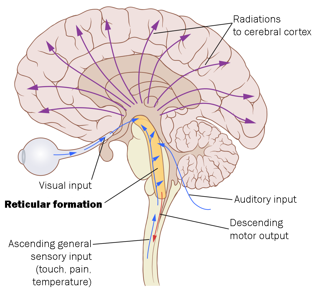 A loosely organized group of cells forms the central core of the brainstem, extending through the medulla, pons, and midbrain. This core group of neurons appears net-like in some staining methods, so it has the name of reticular formation (Latin: reticulum, “little net,” “hair-net”).
The reticular formation is the alarm clock of the brain. If you are sleeping in your cave and a sabre-toothed tiger nibbles on your toe, your reticular formation will wake you up and get you moving. Conversely, if the reticular formation is damaged, as in coma, patients cannot be aroused and seem to always be in a sleep-like state.
The reticular formation takes in information from many sensory systems, and then after processing, sends information over a wide area of cerebral cortex to keep all areas of the brain active.
Media Attributions
- U11-117 Limbic System © BruceBlaus is licensed under a CC BY (Attribution) license
- U11-117a New Brain Regions Involved in Mental Illness © National Institute of Mental Health, National Institutes of Health is licensed under a Public Domain license
- U11-118 Hypothalamus Pituitary Complex © Betts, J. Gordon; Young, Kelly A.; Wise, James A.; Johnson, Eddie; Poe, Brandon; Kruse, Dean H. Korol, Oksana; Johnson, Jody E.; Womble, Mark & DeSaix, Peter is licensed under a CC BY (Attribution) license
- U11-119 Hypothalamus © National Institutes of Health is licensed under a Public Domain license
- U11-120 Hypothalamo Pituitary Adrenal (HPA) Stress Axis © Murgatroyd, Chris and Spengler, Dietmer is licensed under a CC BY (Attribution) license
- U11-121 Reticular Formation © Lynch, Patrick J. adapted by Jim Hutchins is licensed under a CC BY-SA (Attribution ShareAlike) license

_stress_axis.jpg)